


We report the case of a 78-year-old woman with a history of hypertension, deep vein thrombosis, valvular heart disease treated with coumarin, osteoporosis, and overlap syndrome. In addition, she had diagnostic findings consistent with systemic lupus erythematosus (pleuropericarditis, lupus erythematosus panniculitis, subacute cutaneous lupus erythematosus, malar rash, oral ulcers, arthritis, leukopenia and thrombocytopenia, meningitis, and positive tests for antinuclear antibodies and antiribonucleoprotein antibodies) as well as with scleroderma (sclerodactyly, severe Raynaud syndrome, esophageal disease, interstitial pulmonary disease, and a positive test for anticentromere antibodies). The patient presented to our dermatology service in 2002 because of bouts of erythema and pain in her right leg. Biopsy findings at that time were reported as consistent with lupus erythematosus panniculitis and dermal sclerosis. In 2008 she began to develop ulcers above the malleoli of her right leg. The lesions were very painful and frequently infected. Results of another biopsy, performed in 2009, were consistent with calcified scleroderma. The patient's calcium-phosphorus product was normal, and a radiograph of her leg revealed subcutaneous calcification. All these findings were consistent with dystrophic calcification. We initially treated the patient with oral corticosteroids, with courses of antibiotics added whenever infection occurred.
In November 2009, we began treatment with bosentan, but discontinued treatment owing to poor tolerance. In December 2009 we began topical treatment with sodium thiosulfate and acetic acid. The patient's condition initially improved, and Pseudomonas infections stopped. Between January and June 2010, we added sildenafil to treat the patient for digital ulcers, which resolved. The leg ulcer secondary to dystrophic calcification remained unaltered. In June 2010, the patient's calcified deposits were excised and the defect was covered with a skin graft. The ulcer recurred 4 months later and diltiazem was prescribed. Initial improvement was considerable, but a subsequent recurrence failed to respond both to doxycycline and to several courses of corticosteroids.
Owing to this poor response, in July 2012 we decided to start treatment in our rehabilitation service using unfocused shock waves at an intensity of 0.1mJ/mm2 and a frequency of 360 pulses per minute, with 550 pulses administered to the proximal area and 400 to the distal area. Slight adjustments were made as the size of the ulcer changed. Sessions lasted approximately 5minutes each and were conducted every 2 weeks. The patient tolerated this treatment very well and experienced no adverse effects. Her symptoms improved considerably from the first session onwards; pain was reduced and lesions became progressively smaller until they were practically epithelialized (Figs. 1 and 2).


Dystrophic calcification is the term used for the formation of insoluble calcified deposits in the skin and soft tissues (calcinosis cutis) owing to tissue damage in individuals with normal calcium and phosphorus serum levels.1 It is frequently, but not exclusively, associated with autoimmune diseases of connective tissue, particularly dermatomyositis (20%-70%) and the localized form of systemic scleroderma (25%), in which it occurs in the areas most severely affected by sclerosis and ischemia.2 It is also a typical finding in biopsies for long-standing lupus erythematosus panniculitis.3
Manifestations range from radiologic findings to highly painful chronic nodules and ulcers that frequently become infected and impair quality of life. These ulcers are challenging to manage, and no treatment has yet been shown to be universally effective. Treatments attempted include antiinflammatory intralesional corticosteroids, calcium antagonists (diltiazem), colchicine, minocycline, bisphosphonates, warfarin, intravenous immunoglobulin, probenecid, aluminum hydroxide, ceftriaxone, topical sodium thiosulfate, surgery, and erbium:YAG or carbon dioxide laser therapy.4
Extracorporeal shock waves have been used in urology since 1980 and in trauma and orthopedics since 1988. Calcific tendinitis of the shoulder is one of the indications. Shock wave treatment consists of high-pressure acoustic pulses generated with an impulse faster than the speed at which the sound waves propagate within a given medium; this generally involves an electric discharge in a watery medium. Several types of reflector focus the waves according to the treatment objective, giving rise to either high-density focused wave fronts (for calculi) or low-density unfocused wave fronts (for soft tissue).5 Low-density waves act on cell-surface mechanoreceptors and activate angiogenesis as well as the migration and differentiation of cells with high regenerative potential. They also stimulate sensory nerve fibers and nociceptors, a fact that may explain their analgesic effect.6
Our literature searches have found 9 published cases of patients with ulcers secondary to dystrophic calcification who were treated with focused waves (for calculi)7,8: 4 had chronic venous insufficiency, 4 had scleroderma, and 1 had dermatomyositis. Additionally, there was 1 patient who had dermatomyositis but no ulcers.9 All these patients experienced significant decreases in ulcer size and pain after 2 to 3 sessions, across all conditions.
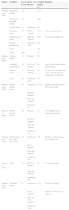
In our patient's case we opted for unfocused low-density waves using equipment designed for soft tissue.10 This therapy has been used for different types of ulcers since 2007, with promising results (Table 1).5
Published Uses of Shock Waves to Heal Ulcersa
| Author | Condition | No. of Ulcers | Shock wave therapy | Complete healing (%) | Remarks |
|---|---|---|---|---|---|
| Schaden et al., 2007 | Anomalous healing | 82 | 75.6 | ||
| Necrosis of traumatic lesions | 67 | 86.6 | |||
| Venous stasis | 25 | Unfocused | 36 | ||
| Decubitus ulcers | 14 | ED: 0.1mJ/mm2 | 71.4 | 1/3 were acute ulcers. | |
| Other pressure ulcers | 7 | Pulse no.: 100/cm2 | 85.7 | No long-term follow-up. | |
| Arterial insufficiency | 6 | Frequency: 5Hz | 66.7 | ||
| Burns | 7 | 100 | |||
| Total | 208 | 75 | |||
| Saggini et al., 2008 | Venous ulcers | 12 | Focused | 36 | |
| Traumatic wound ulcers | 16 | ED: 0.037mJ/mm2 | 69 | Only 1/10 of control group ulcers healed. | |
| Diabetic ulcers | 4 | Pulse no.: 100/cm2 | 25 | Wound size was reduced in every case and pain was significantly diminished. | |
| Total | 32 | Frequency: 4Hz | 50 | ||
| Moretti et al., 2009 | Plantar diabetic ulcers | 30 | Unfocused | 53.35 | Healing was faster than in the control group. |
| ED: 0.03mJ/mm2 | |||||
| Pulse no.: 100/cm2 | |||||
| Frequency: NA | |||||
| Wang et al., 2009 | Plantar diabetic ulcers | 36 | Focused | 31 | 22% healing rate in hyperbaric chamber. |
| ED: 0.11mJ/mm2 | |||||
| Pulse no.: 100/cm2 | |||||
| Frequency: NA | |||||
| Ottoman et al., 2010 | Full-thickness skin graft donor areas | 28 | Unfocused | 100 | Healing was faster than in the control group. |
| ED: 0.1mJ/mm2 | |||||
| Pulse no.: 100/cm2 | |||||
| Frequency: NA | |||||
| Arno et al., 2010 | Burns | 15 | ED: 0.15mJ/mm2 | 80 | No control group. |
| Pulse no.: 500/cm2 | |||||
| Frequency: NA | |||||
| Larking et al., 2010 | Decubitus ulcers | 9 | Unfocused | 55.5 | Crossover design. |
| ED: 0.1mJ/mm2 | Shock wave therapy was superior. | ||||
| Pulse no.: 200+100/cm2 | |||||
| Frequency: 5Hz |
Abbreviations: ED, energy density; NA, not available.
To our knowledge, ours is the first case of an ulcer caused by dystrophic calcification and treated using unfocused shock waves, with excellent results. We wish to highlight the effectiveness of this treatment in terms of pain reduction and epithelialization, and underscore its ease of application, safety, and tolerability.
Please cite this article as: Delgado-Márquez AM, Carmona M, Vanaclocha F, Postigo C. Eficacia del tratamiento con ondas de choque en las úlceras por calcinosis distrófica. Actas Dermosifiliogr. 2015;106:140–143.


