


A 76-year-old man presented with a slow-growing lesion that had appeared 4 years earlier on the great toe of his left foot. The patient reported recurring ingrowth of the nail on the same toe prior to the appearance of the lesion, as well as occasional episodes of self-limited bleeding. The lesion was not painful, except when the nail was ingrown or after minor trauma.
Physical ExaminationThe lesion was a partially eroded polylobulated botryomycosis-like tumor with an indurated base and softer surface. It measured approximately 3×3cm and covered the mid-distal part of the great toe of the left foot, displacing the nail (Fig. 1).
Histopathology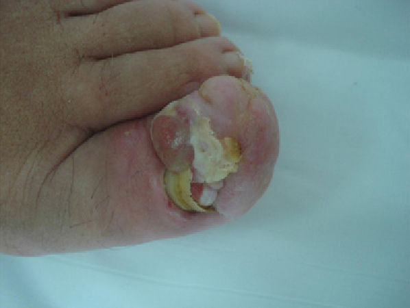
Histopathological findings showed anastomosing cords of epithelial cells connected to the epidermis and embedded in a fibrovascular stroma. The epithelial cords contained luminal cells of different sizes (Figs. 2 and 3).
Additional Tests

Magnetic resonance imaging disclosed a 3.5×3×2cm multilobulated subungual nodule confined to the soft tissue, with no sign of bone destruction.
What Is Your Diagnosis?
DiagnosisEccrine syringofibroadenoma.
Clinical Course and TreatmentThe lesion was excised, resulting in definitive cure. No recurrence has been observed to date.
CommentEccrine syringofibroadenoma (ESFA) is a rare tumor that was described by Mascaró in 1963.1 Clinical presentation varies, ranging from solitary lesions to multiple papules and nodules. The clinical classification Starink2 proposed in 1997 named 4 subtypes: solitary ESFA, multiple ESFA as a marker of the Schöpf syndrome (ectodermal dysplasia), multiple ESFA with no associated skin findings, and nonhereditary unilateral linear ESFA (also referred to as unilateral linear nevoid syringofibroadenoma). Solitary ESFA, the most common type, usually forms in the distal region of the limbs. Histologically, the tumor is composed of thin cords and strands of epithelial cells in a network connected to the epidermis. Cells are paler than epidermal keratinocytes and luminal cells surrounded by an eosinophilic cuticle can often be found within cords. A highly fibrovascular stroma lies between the strands and the cords.3 The histologic, ultrastructural, and immunohistochemical features reported in the literature suggest that this tumor has an acrosyringeal and eccrine intradermal ductal nature.4–6
The pathogenesis of solitary ESFA is poorly understood. Some authors suggest that it is not a true benign neoplasm of the eccrine glands but rather reactive hyperplasia of the eccrine ducts as a response to repeated tissue damage (chronic ulcers, chronic lymphedema, burns, nail trauma, etc.).4 In this context, a new reactive subtype of solitary ESFA has been proposed.4–6 Our case, in which the lesion developed secondary to repeated ingrowth of the toenail, could be considered a reactive ESFA. Recent reports have described cases of solitary ESFA associated with squamous cell carcinoma and the malignant transformation of solitary ESFA. Histologically, this diagnosis must be differentiated from fibroepithelioma of Pinkus, tumors of the follicular infundibulum, and squamous cell carcinoma. Solitary ESFA is treated according to the number, site, and resectability of the lesions, and simple excision is the treatment of choice in solitary tumors.6
Please cite this article as: Bernat-García J, et al. Tumoración en el primer dedo del pie izquierdo. Actas Dermosifiliogr. 2013;104:523–4.


