


The 2 patients presented pigmented lesions with a similar clinical (Fig. 1) and dermoscopic (Fig. 2) appearance. However, detailed evaluation of the dermoscopic images provides clues to the diagnosis in each case. Both tumors are formed of multiple, dark, round or oval structures in apposition. In my opinion, one of the diagnostic keys is to determine whether or not these structures are dark lacunae. Dark lacunae are multiple, well-defined, round or oval structures that are dark blue, violaceous, or black. Histologically, these structures are seen to be partially or completely thrombosed dilated blood vessels in the dermis, and their presence is highly suggestive of solitary angiokeratoma (sensitivity, 93.8%; specificity, 99.1%).1 An important negative finding of lacunae is that there are no vascular structures in their interior.

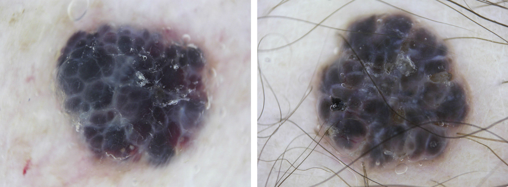
In the dermoscopic image of the first patient there are multiple, well-defined, round, oval, or occasionally polygonal structures (slightly out of focus on the right side of the image) that are violaceous or dark blue and do not contain blood vessels. These structures are dark lacunae and the diagnosis suggested by the dermoscopic image is solitary angiokeratoma.
In the dermoscopic image of the second patient the structures are slightly different. They are multiple, round or oval structures but they are poorly defined and are grayish-blue in color. Furthermore, a vascular structure, in perfect focus, formed of a thick central blood vessel that branches into smaller vessels, is visible in one of the structures at the left inferior pole of the nodule. Based on the comments above, these structures cannot be called lacunae. However, their description does match that of a recognized dermoscopic feature: large blue-gray ovoid nests. This is 98% specific for basal cell carcinoma.2 Thus, if no criteria for a melanocytic lesion are observed and we recognize that the vascular structure described is a tree-like or arborizing telangiectasia, which has a positive predictive value of 94% for basal cell carcinoma,3 we should make a dermoscopic diagnosis of pigmented basal cell carcinoma.
Conflicts of InterestThe author declares that he has no conflicts of interest.
Please cite this article as: Zaballos Diego P. Encuentra las diferencias entre 2 pápulas pigmentadas. Actas Dermosifiliogr. 2013;104:719–20.


