


The patient, a 14-year-old boy with no relevant personal or family history, presented with asymptomatic lesions on both hands that had first appeared approximately 1 year earlier. He had received treatment with topical corticosteroids on several occasions. The lesions had improved somewhat during each round of treatment, only to worsen again after the medication was withdrawn.
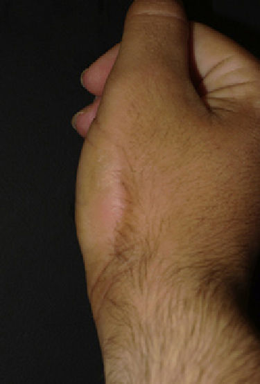
Physical ExaminationPhysical examination revealed yellowish-white keratotic plaques with well-defined borders on the dorsum of the right hand and on the lateral sides of the proximal phalanges of both hands (Fig. 1, A and B). A larger plaque with similar characteristics was located on the right thenar eminence, stretching around to the lateral edge of the hand (Fig. 2). The patient had no other lesions.
Histopathology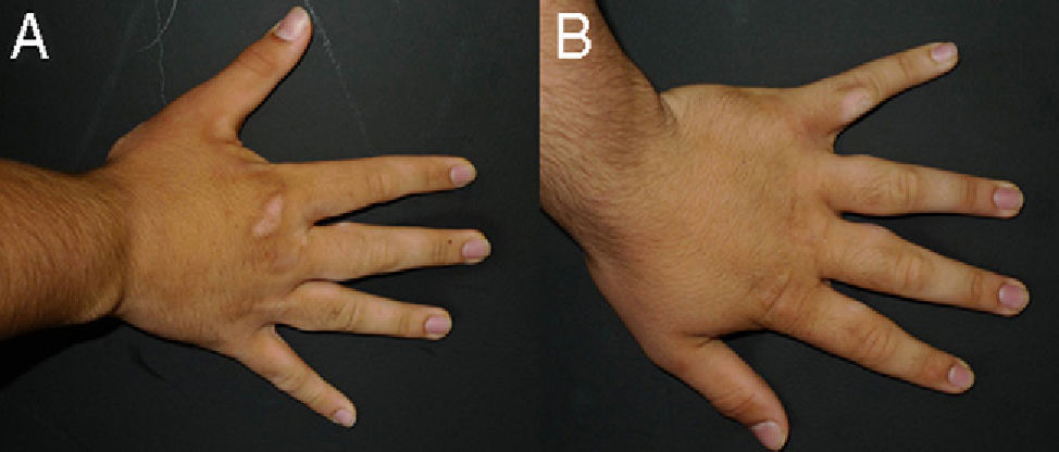
Histopathology revealed marked orthokeratotic hyperkeratosis and mild epidermal acanthosis (Fig. 3A). Orcein staining revealed a reduction (Fig. 3B) and fragmentation (Fig. 3C) of the elastic fibers in the dermis.

What Is Your Diagnosis?
DiagnosisAcrokeratoelastoidosis of Costa.
Clinical Course and TreatmentDue to the benign nature of the condition and the absence of symptoms, the patient was given no treatment.
CommentFirst described by Costa1 in 1953, acrokeratoelastoidosis (AKE) is a rare entity that can be either sporadic or familial. Autosomal dominant and autosomal recessive inheritance patterns have been described, as well as a possible linkage to chromosome 2.2 In most cases, onset occurs before the age of 20 years,3 and the disease exhibits no clear predilection for race or sex.4
The lesions are small, translucent, yellowish-white papules measuring 2 to 4mm. They can be round or polygonal, have a flat or umbilicated surface, and are often keratotic. The most characteristic locations are the thenar and hypothenar eminences and the dorsal and lateral aspects of the fingers and toes.2-5 The lesions typically occur across the areas where the dorsal and plantar or the dorsal and palmar surfaces meet.3 On the dorsa of the feet, the papules tend to merge into plaques5 that can extend as far as the anterior aspect of the legs; lesions rarely occur on the soles of the feet.4 The lesions are usually asymptomatic, although pruritus2 and pain caused by fissuring have been reported.6 Hyperhidrosis is a common but variable finding.1,4
Histology reveals epidermal acanthosis with orthokeratotic hyperkeratosis. In the dermis, a reduction and fragmentation of elastic fibers (elastorrhexis) is observed.2-4 In some cases, these changes in the skin can only be observed by electron microscopy.5
The cause of AKE is unknown,2,4 but electron microscopic studies of patients with AKE have revealed fibroblasts containing dense cytoplasmic granules and an absence of extracellular fibers. These findings support the hypothesis that the underlying disease process is the inhibition of elastic fiber synthesis by fibroblasts.5
Key conditions that should be considered in the differential diagnosis include other marginal acrokeratodermas such as focal acral hyperkeratosis, degenerative collagenous plaques of the hands, keratoelastoidosis marginalis of the hands, and lenticular acral keratosis in laundry workers. Diagnosis is established on the basis of clinical findings and histologic examination of the lesions.2,4
Treatment is generally unnecessary, as the lesions are usually asymptomatic. Of the treatments used to date—dapsone, methotrexate, topical salicylic acid, topical capsaicin,6 liquid nitrogen, oral and topical corticosteroids, and erbium:YAG laser treatment—oral retinoid treatment is the only one likely to lead to any improvement; it should be noted, however, that the lesions recur after discontinuation of the treatment.2,4
Please cite this article as: Alonso-González J, et al. Placas queratósicas en manos. Actas Dermosifiliogr.2012;103:327-8.


