


Herpes zoster (shingles) is rare in children with no history of chickenpox but can occur when their mothers are infected with the virus during pregnancy. In such cases, herpes zoster generally presents after the first year of life after maternal immunoglobulin (Ig) G antibodies have disappeared from the child's blood. Disseminated pediatric herpes zoster is an unusual condition.
We present the case of a 2-year-old child who was treated at the emergency department of our hospital in July 2011 with an acute and painful rash distributed metamerically in the left thoracic region. The patient was diagnosed with herpes zoster and treated with hot dry compresses. The medical history showed that the mother had contracted chickenpox at pregnancy week 21. In all other respects, the patient was a healthy child who was not taking any medications. She had an up-to-date vaccination schedule, but had not been vaccinated against the varicella zoster virus. Five days later, the patient was brought back to the emergency department because the rash had spread and there was associated pain, fever, and anorexia.
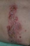
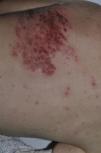
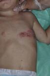
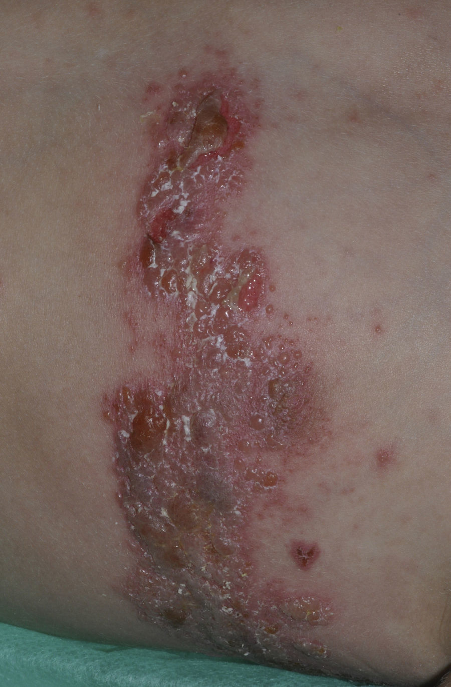
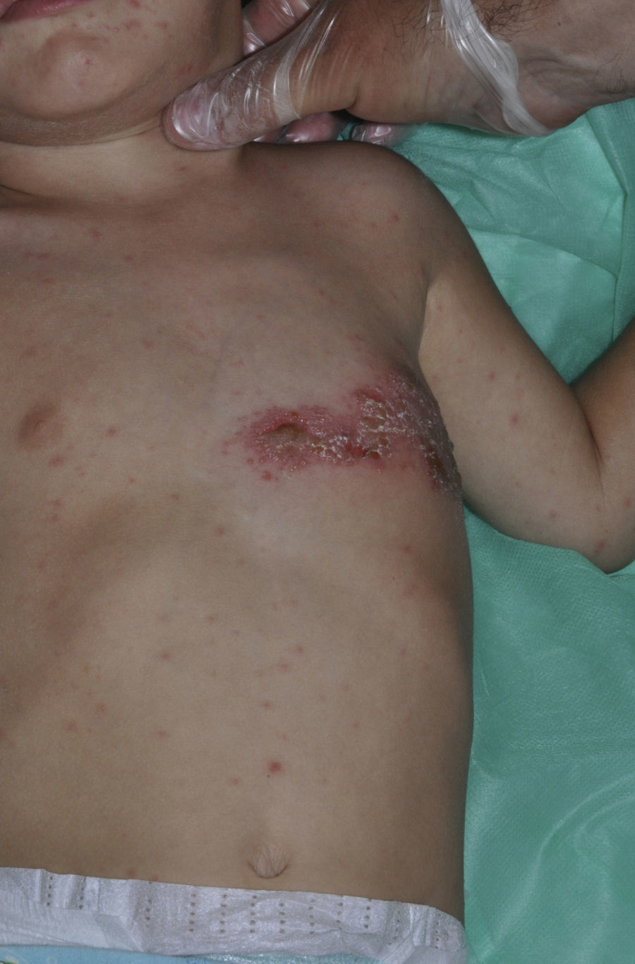
Examination of the skin revealed a rash consisting of clustered vesicles (some containing blood) on an erythematous base, distributed metamerically following dermatomes T4 and T5 (Fig. 1). We also observed a generalized rash made up of hundreds of isolated vesicles all over the body (Figs. 2 and 3). No palpable lymph nodes were detected.

The child was hospitalized with a diagnosis of disseminated pediatric herpes zoster and treated with intravenous acyclovir at a dosage of 10mg/kg every 8hours for 5 days. She also received intravenous analgesia, and the rash was treated with topical potassium permanganate. Laboratory test results revealed mild iron deficiency (15 mcg/dL), no anemia, and high titers of IgG herpes zoster antibodies. The results of other immune studies, including serology for cytomegalovirus and human immunodeficiency virus (HIV) were normal or negative. No signs or symptoms of visceral involvement were detected. After 5 days of treatment, the patient's symptoms improved and she was discharged; oral iron supplementation was prescribed.
The impact on the fetus of maternal varicella infection during pregnancy depends on when the infection occurs.1–3 If the mother is infected before week 20, there is a high risk of miscarriage, although in some cases such infection can lead to congenital varicella syndrome. The clinical effects of congenital varicella syndrome are usually severe and include pitted and pigmented dermatomal skin scarring, cataracts, chorioretinitis, Horner syndrome, limb hypoplasia, cortical atrophy, seizures, and mental retardation.3
If maternal infection occurs between weeks 21 and 28, in most cases the child will not have symptoms at birth because the infection will be controlled by maternal IgG antibodies.2 The child may develop herpes zoster later in life once these antibodies disappear (which occurs after 1 or 2 years); this was the case in our patient.1,3
If maternal infection is perinatal (within 5 days prior to delivery), there is a high risk of neonatal chickenpox, which may progress mildly if it presents with skin involvement alone. However, complications are more common at this age due to the poorly developed immune system of the neonate and may include pneumonia, hepatitis, and meningoencephalitis. The risk of neonatal chickenpox increases in premature and post-term infants and in cases of perinatal infection because in these conditions the child receives less protection from the maternal IgG antibodies and their own production of IgG antibodies is low.
As with adult herpes zoster, pediatric herpes zoster is caused by reactivation of latent varicella-zoster virus in nerve ganglia. Disseminated pediatric herpes zoster is a rare condition and, as with the adult form, it is more often found in immunosuppressed patients (HIV, lymphomas, immunosuppressant therapy), who tend to have visceral involvement.4 It is therefore important to rule out underlying immunosuppression and visceral involvement in patients with disseminated forms of the disease. In cases with only skin involvement, the prognosis is good and treatment with oral acyclovir is recommended at a dosage of 20 to 30mg/kg/d, divided into 5 doses, each administered at 4-hour intervals, with a maximum dosage of 800mg/d for 7 days. Higher dosages are only used in cases of visceral involvement. For cases involving acyclovir resistance, the treatment of choice is intravenous foscarnet.
In summary, we present the case of a 2-year-old immunocompetent patient who developed disseminated pediatric herpes zoster; the only medical history of interest was maternal varicella infection during pregnancy. Disseminated pediatric herpes zoster is a rare condition that usually affects immunosuppressed individuals and is generally associated with visceral involvement.4 Nevertheless, 5 other cases of patients with disseminated disease confined to the skin have been published1,5,6; 2 of the patients also presented iron deficiency with no anemia.4 However, no association between iron deficiency and pediatric herpes zoster has yet been established.
Please cite this article as: Ortiz-Brugués A, et al. Herpes zóster diseminado infantil. Actas Dermosifiliogr. 2013;104:441-2.


