


Rosai-Dorfman disease is a benign histiocytic proliferative disorder of unknown etiology. The presenting sign is usually asymptomatic massive lymphadenopathy. Exclusively cutaneous cases of Rosai-Dorfman disease are rare. We present a case report of the disease and describe its dermoscopic features.
A 29-year old man with no relevant medical or surgical history was seen for a lesion that had been growing progressively for several years in the left pectoral region. A biopsy report from another hospital indicated diagnosis of atypical xanthogranuloma.
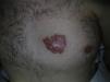
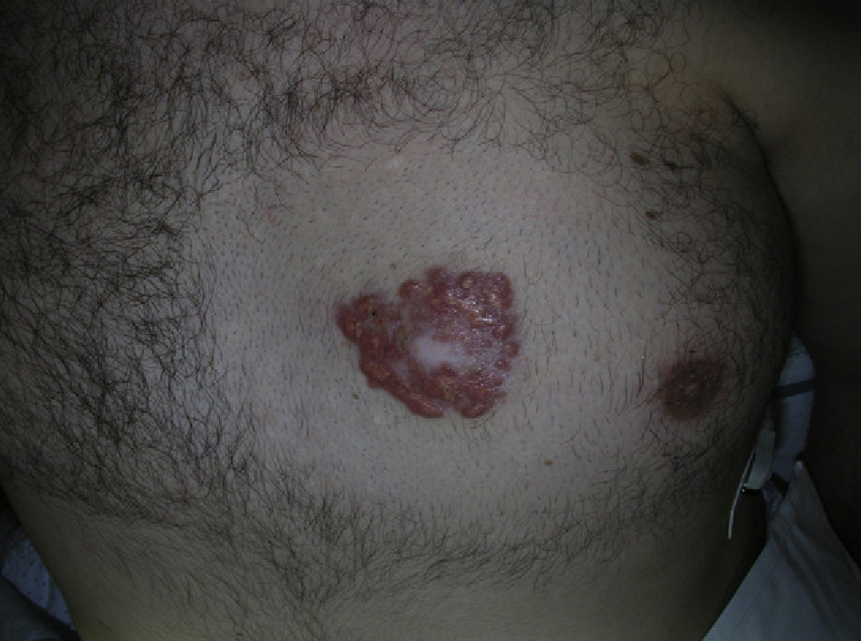
The physical examination showed an infiltrated plaque with a heterogeneous surface and well-defined borders. Its longest diameter was 4.5×4cm and it had an erythematous-orange appearance, a central whitish area, and multiple yellowish structures at the edges (Fig. 1).
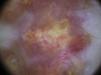
The dermoscopic image showed a milky-red area in the center of the lesion, whereas the outer part had multiple yellowish homogeneous areas of different sizes with irregular borders, surrounded by large telangiectatic vessels on a red-orange background (Fig. 2). Given the progressive growth of the lesion and the discomfort it caused the patient, radical excision was performed.

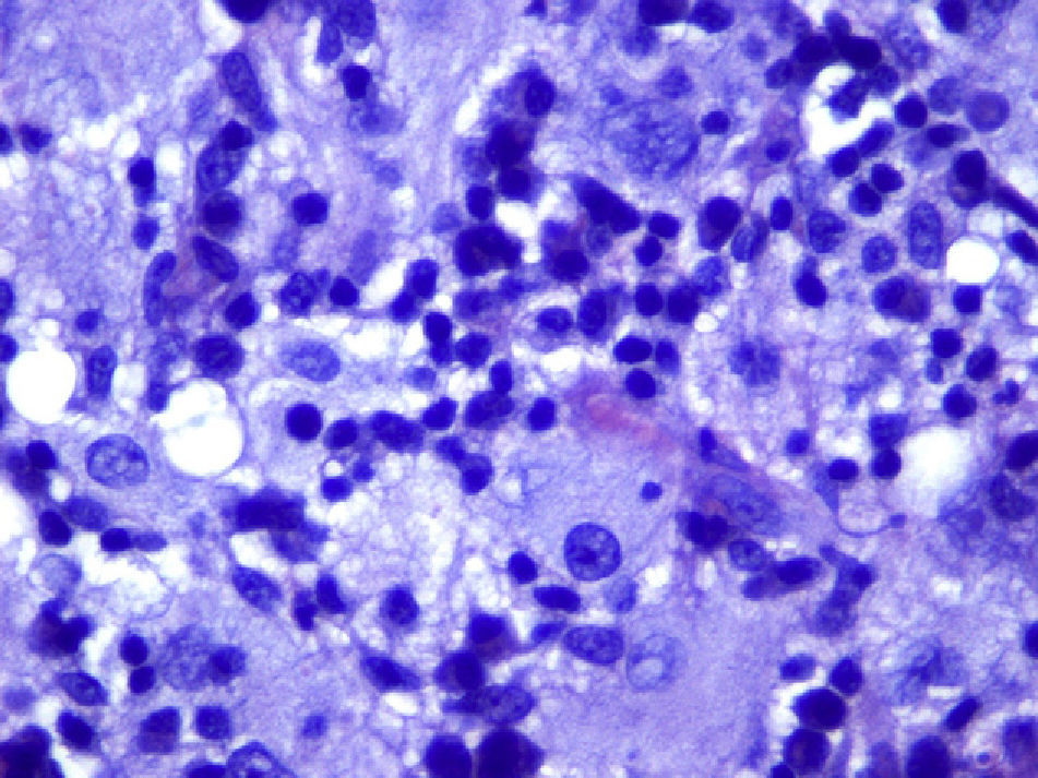
Histologic examination revealed a poorly-defined dermal proliferation of large histiocytes. extending downwards into the subcutaneous adipose tissue. The histiocytes had large eosinophilic vacuolated cytoplasm and round homogeneous nuclei and some contained intact lymphocytes (Fig. 3). In addition to the histiocytes, there was abundant inflammatory infiltrate composed mainly of plasma cells, with lymphocytes, giant multinucleated cells, and xanthomatous histiocytes with small nuclei. A prominent fibrous stroma was seen with a highly vascularized storiform pattern. Immunohistochemical staining was positive for S-100 and CD68 and negative for CD1a. The presence of emperipolesis and the immunohistochemical profile pointed to diagnosis of a cutaneous variant of Rosai-Dorfman disease. No hematologic abnormalities, lymph node involvement, or abnormalities in other organs were observed in the staging study.
Rosai-Dorfman disease, or massively enlarged lymph nodes with sinus histiocytosis, is a histiocytic proliferation in which approximately 40% of the patients have extranodal involvement.1,2 The skin is the most frequently affected organ. Purely cutaneous forms of Rosai-Dorfman disease are rare. Skin lesions are nonspecific and may take the form of solitary or multiple lesions of different sizes and morphologies. They can present on any part of the body and the clinical differential diagnosis includes a broad range of conditions including panniculitis, vasculitis, acne vulgaris, suppurative hidradenitis, granuloma annulare, and sarcoidosis, as well as other histiocytoses.3 Kong et al.4 proposed a classification based on the morphologic features of 39 lesions. Papulonodular lesions were the most common form (accounting for almost 80%) followed by the infiltrated plaque type (12.5%) and the tumor type (7.7%). The histologic findings in cutaneous lesions are similar to those found in lymph tissue. The main finding is a dense infiltrate of large histiocytes and a large pale cytoplasm with rounded nuclei. The cytoplasm contains intact leukocytes, usually lymphocytes, a phenomenon known as lymphophagocytosis or emperipolesis. Typically, these histiocytes are positive for S-100 and negative for CD1a, and can be either positive or negative for CD68. The immunohistochemical profile is essential for histologic diagnosis, as fibrosis, vascular proliferation, lymphoid clusters, foam cells, and multinucleated Touton giant cells may or may not be present, and so confusion with other histiocytic processes, and with juvenile xanthogranuloma in particular, is possible.The dermoscopic features of Rosai-Dorfman disease have not been widely reported in the literature.5,6 Rodríguez Blanco et al.6 reported a case of Rosai-Dorfman disease on the sole, characterized by cotton-like ovoid structures on an erythematous background in the dermoscopic image. In contrast, the dermoscopic features in our case were similar to those described for juvenile xanthogranuloma, that is, a yellow-orange homogeneous central area and a somewhat more erythematous peripheral area. This is known as the setting sun feature.7 The presence of clouds of pale yellow globules is considered indicative of xanthomatous histiocytes in the superficial dermis.8 The presence of comma vessels,9 arborizing telangiectasia,10 and whitish linear projections has also been reported, particularly in advanced cases of juvenile xanthogranuloma. The differential dermoscopic diagnosis should be performed with solitary yellow lesions, such as juvenile xanthogranuloma, organoid nevus or sebaceous nevus, xanthomatous dermatofibroma, and solitary reticulohistiocytoma.9–11
The lesion we describe was large, morphologically irregular, with dermoscopic features similar to those described for juvenile xanthogranuloma or solitary reticulohistiocytoma, although our lesion had a wider range of coloration, with a milky-red central zone, multiple yellow clouds surrounded by arborizing vessels, and an erythematous-orange peripheral area.
In conclusion, cutaneous Rosai-Dorfman disease is rare and lacks specific clinical characteristics. It should be suspected when an infiltrate of xanthomatous histiocytes is observed, particularly if these histiocytes contain intact lymphocytes. Given that the differential diagnosis of this disease can be difficult, dermoscopy can be useful, particularly when features with a yellow coloration are present, as in the case presented here.
Please cite this article as: Avilés-Izquierdo JA, et al. Características dermatoscópicas de la enfermedad de Rosai-Dorfman cutánea. Actas Dermosifiliogr. 2012;103:446-8.


