array:24 [
  "pii" => "S1578219013002163"
  "issn" => "15782190"
  "doi" => "10.1016/j.adengl.2012.08.022"
  "estado" => "S300"
  "fechaPublicacion" => "2013-11-01"
  "aid" => "731"
  "copyright" => "Elsevier España, S.L. and AEDV"
  "copyrightAnyo" => "2012"
  "documento" => "simple-article"
  "crossmark" => 0
  "subdocumento" => "cor"
  "cita" => "Actas Dermosifiliogr. 2013;104:826-8"
  "abierto" => array:3 [
    "ES" => true
    "ES2" => true
    "LATM" => true
  ]
  "gratuito" => true
  "lecturas" => array:2 [
    "total" => 3435
    "formatos" => array:3 [
      "EPUB" => 43
      "HTML" => 2916
      "PDF" => 476
    ]
  ]
  "Traduccion" => array:1 [
    "es" => array:19 [
      "pii" => "S0001731012004486"
      "issn" => "00017310"
      "doi" => "10.1016/j.ad.2012.08.002"
      "estado" => "S300"
      "fechaPublicacion" => "2013-11-01"
      "aid" => "731"
      "copyright" => "Elsevier España, S.L. y AEDV"
      "documento" => "simple-article"
      "crossmark" => 0
      "subdocumento" => "cor"
      "cita" => "Actas Dermosifiliogr. 2013;104:826-8"
      "abierto" => array:3 [
        "ES" => true
        "ES2" => true
        "LATM" => true
      ]
      "gratuito" => true
      "lecturas" => array:2 [
        "total" => 5234
        "formatos" => array:3 [
          "EPUB" => 2
          "HTML" => 4189
          "PDF" => 1043
        ]
      ]
      "es" => array:11 [
        "idiomaDefecto" => true
        "cabecera" => "<span class="elsevierStyleTextfn">Carta cient&#237;fico-cl&#237;nica</span>"
        "titulo" => "Dermatoscopia de tumores apocrinos&#58; presentaci&#243;n de 4 casos"
        "tienePdf" => "es"
        "tieneTextoCompleto" => "es"
        "paginas" => array:1 [
          0 => array:2 [
            "paginaInicial" => "826"
            "paginaFinal" => "828"
          ]
        ]
        "titulosAlternativos" => array:1 [
          "en" => array:1 [
            "titulo" => "Dermatoscopy of Apocrine Tumors&#58; Report of 4 Cases"
          ]
        ]
        "contieneTextoCompleto" => array:1 [
          "es" => true
        ]
        "contienePdf" => array:1 [
          "es" => true
        ]
        "resumenGrafico" => array:2 [
          "original" => 0
          "multimedia" => array:7 [
            "identificador" => "fig0015"
            "etiqueta" => "Figura 3"
            "tipo" => "MULTIMEDIAFIGURA"
            "mostrarFloat" => true
            "mostrarDisplay" => false
            "figura" => array:1 [
              0 => array:4 [
                "imagen" => "gr3.jpeg"
                "Alto" => 1170
                "Ancho" => 2647
                "Tamanyo" => 449117
              ]
            ]
            "descripcion" => array:1 [
              "es" => "<p id="spar0015" class="elsevierStyleSimplePara elsevierViewall">Im&#225;genes histol&#243;gicas &#40;hematoxilina-eosina &#215;20&#41;&#46; Quiste en la dermis revestido por una doble capa de c&#233;lulas cuboideas &#40;a y b&#41;&#46; Tumor bien circunscrito constituido por c&#233;lulas de citoplasma amplio y eosin&#243;filo y otras de aspecto m&#225;s basaloide&#46; Estructuras qu&#237;sticas en el centro de la lesi&#243;n &#40;c&#41;&#46;</p>"
            ]
          ]
        ]
        "autores" => array:1 [
          0 => array:2 [
            "autoresLista" => "D&#46;E&#46; Cieza-D&#237;az, J&#46;A&#46; Avil&#233;s-Izquierdo, C&#46; Ciudad-Blanco, V&#46; Parra-Blanco"
            "autores" => array:4 [
              0 => array:2 [
                "nombre" => "D&#46;E&#46;"
                "apellidos" => "Cieza-D&#237;az"
              ]
              1 => array:2 [
                "nombre" => "J&#46;A&#46;"
                "apellidos" => "Avil&#233;s-Izquierdo"
              ]
              2 => array:2 [
                "nombre" => "C&#46;"
                "apellidos" => "Ciudad-Blanco"
              ]
              3 => array:2 [
                "nombre" => "V&#46;"
                "apellidos" => "Parra-Blanco"
              ]
            ]
          ]
        ]
      ]
      "idiomaDefecto" => "es"
      "Traduccion" => array:1 [
        "en" => array:9 [
          "pii" => "S1578219013002163"
          "doi" => "10.1016/j.adengl.2012.08.022"
          "estado" => "S300"
          "subdocumento" => ""
          "abierto" => array:3 [
            "ES" => true
            "ES2" => true
            "LATM" => true
          ]
          "gratuito" => true
          "lecturas" => array:1 [
            "total" => 0
          ]
          "idiomaDefecto" => "en"
          "EPUB" => "https://multimedia.elsevier.es/PublicationsMultimediaV1/item/epub/S1578219013002163?idApp=UINPBA000044"
        ]
      ]
      "EPUB" => "https://multimedia.elsevier.es/PublicationsMultimediaV1/item/epub/S0001731012004486?idApp=UINPBA000044"
      "url" => "/00017310/0000010400000009/v1_201310240057/S0001731012004486/v1_201310240057/es/main.assets"
    ]
  ]
  "itemSiguiente" => array:19 [
    "pii" => "S1578219013002175"
    "issn" => "15782190"
    "doi" => "10.1016/j.adengl.2012.08.023"
    "estado" => "S300"
    "fechaPublicacion" => "2013-11-01"
    "aid" => "741"
    "copyright" => "Elsevier Espa&#241;a&#44; S&#46;L&#46; and AEDV"
    "documento" => "simple-article"
    "crossmark" => 0
    "subdocumento" => "cor"
    "cita" => "Actas Dermosifiliogr. 2013;104:829-30"
    "abierto" => array:3 [
      "ES" => true
      "ES2" => true
      "LATM" => true
    ]
    "gratuito" => true
    "lecturas" => array:2 [
      "total" => 2096
      "formatos" => array:3 [
        "EPUB" => 37
        "HTML" => 1623
        "PDF" => 436
      ]
    ]
    "en" => array:11 [
      "idiomaDefecto" => true
      "cabecera" => "<span class="elsevierStyleTextfn">Case and Research Letters</span>"
      "titulo" => "Pityriasis Lichenoides in a 9-Month-Old Boy"
      "tienePdf" => "en"
      "tieneTextoCompleto" => "en"
      "paginas" => array:1 [
        0 => array:2 [
          "paginaInicial" => "829"
          "paginaFinal" => "830"
        ]
      ]
      "titulosAlternativos" => array:1 [
        "es" => array:1 [
          "titulo" => "Pitiriasis Liquenoide en un Lactante De 9 Meses"
        ]
      ]
      "contieneTextoCompleto" => array:1 [
        "en" => true
      ]
      "contienePdf" => array:1 [
        "en" => true
      ]
      "resumenGrafico" => array:2 [
        "original" => 0
        "multimedia" => array:7 [
          "identificador" => "fig0010"
          "etiqueta" => "Figure 2"
          "tipo" => "MULTIMEDIAFIGURA"
          "mostrarFloat" => true
          "mostrarDisplay" => false
          "figura" => array:1 [
            0 => array:4 [
              "imagen" => "gr2.jpeg"
              "Alto" => 694
              "Ancho" => 1600
              "Tamanyo" => 389884
            ]
          ]
          "descripcion" => array:1 [
            "en" => "<p id="spar0010" class="elsevierStyleSimplePara elsevierViewall">A&#44; Deep perivascular lymphohistiocytic inflammatory infiltrate with involvement of the dermal-epidermal junction &#40;hematoxylin-eosin&#44; original magnification &#215;10&#41;&#46; B&#44; Detail showing interface alteration&#44; fibrinoid necrosis in the vessel walls&#44; and isolated necrotic keratinocytes &#40;hematoxylin-eosin&#44; original magnification &#215;40&#41;&#46;</p>"
          ]
        ]
      ]
      "autores" => array:1 [
        0 => array:2 [
          "autoresLista" => "M&#46;T&#46; L&#243;pez-Villaescusa, &#193;&#46; Hern&#225;ndez-Mart&#237;n, I&#46; Colmenero, A&#46; Torrelo"
          "autores" => array:4 [
            0 => array:2 [
              "nombre" => "M&#46;T&#46;"
              "apellidos" => "L&#243;pez-Villaescusa"
            ]
            1 => array:2 [
              "nombre" => "&#193;&#46;"
              "apellidos" => "Hern&#225;ndez-Mart&#237;n"
            ]
            2 => array:2 [
              "nombre" => "I&#46;"
              "apellidos" => "Colmenero"
            ]
            3 => array:2 [
              "nombre" => "A&#46;"
              "apellidos" => "Torrelo"
            ]
          ]
        ]
      ]
    ]
    "idiomaDefecto" => "en"
    "Traduccion" => array:1 [
      "es" => array:9 [
        "pii" => "S0001731012004589"
        "doi" => "10.1016/j.ad.2012.08.004"
        "estado" => "S300"
        "subdocumento" => ""
        "abierto" => array:3 [
          "ES" => true
          "ES2" => true
          "LATM" => true
        ]
        "gratuito" => true
        "lecturas" => array:1 [
          "total" => 0
        ]
        "idiomaDefecto" => "es"
        "EPUB" => "https://multimedia.elsevier.es/PublicationsMultimediaV1/item/epub/S0001731012004589?idApp=UINPBA000044"
      ]
    ]
    "EPUB" => "https://multimedia.elsevier.es/PublicationsMultimediaV1/item/epub/S1578219013002175?idApp=UINPBA000044"
    "url" => "/15782190/0000010400000009/v1_201310240051/S1578219013002175/v1_201310240051/en/main.assets"
  ]
  "itemAnterior" => array:19 [
    "pii" => "S1578219013002151"
    "issn" => "15782190"
    "doi" => "10.1016/j.adengl.2012.07.036"
    "estado" => "S300"
    "fechaPublicacion" => "2013-11-01"
    "aid" => "729"
    "copyright" => "Elsevier Espa&#241;a&#44; S&#46;L&#46; and AEDV"
    "documento" => "simple-article"
    "crossmark" => 0
    "subdocumento" => "cor"
    "cita" => "Actas Dermosifiliogr. 2013;104:824-6"
    "abierto" => array:3 [
      "ES" => true
      "ES2" => true
      "LATM" => true
    ]
    "gratuito" => true
    "lecturas" => array:2 [
      "total" => 6247
      "formatos" => array:3 [
        "EPUB" => 35
        "HTML" => 5350
        "PDF" => 862
      ]
    ]
    "en" => array:11 [
      "idiomaDefecto" => true
      "cabecera" => "<span class="elsevierStyleTextfn">Case and Research Letters</span>"
      "titulo" => "Progressive Cribriform and Zosteriform Hyperpigmentation"
      "tienePdf" => "en"
      "tieneTextoCompleto" => "en"
      "paginas" => array:1 [
        0 => array:2 [
          "paginaInicial" => "824"
          "paginaFinal" => "826"
        ]
      ]
      "titulosAlternativos" => array:1 [
        "es" => array:1 [
          "titulo" => "Hiperpigmentaci&#243;n zosteriforme y cribiforme progresiva"
        ]
      ]
      "contieneTextoCompleto" => array:1 [
        "en" => true
      ]
      "contienePdf" => array:1 [
        "en" => true
      ]
      "resumenGrafico" => array:2 [
        "original" => 0
        "multimedia" => array:7 [
          "identificador" => "fig0005"
          "etiqueta" => "Figure 1"
          "tipo" => "MULTIMEDIAFIGURA"
          "mostrarFloat" => true
          "mostrarDisplay" => false
          "figura" => array:1 [
            0 => array:4 [
              "imagen" => "gr1.jpeg"
              "Alto" => 781
              "Ancho" => 900
              "Tamanyo" => 111523
            ]
          ]
          "descripcion" => array:1 [
            "en" => "<p id="spar0005" class="elsevierStyleSimplePara elsevierViewall">Macular lesions of even brown color with a reticulate pattern on normal skin&#44; on the right side of the back&#44; without crossing the midline&#46;</p>"
          ]
        ]
      ]
      "autores" => array:1 [
        0 => array:2 [
          "autoresLista" => "B&#46; Monteagudo, &#193;&#46; Le&#243;n-Mateos, F&#46; Campo-Cerecedo, M&#46; Cabanillas"
          "autores" => array:4 [
            0 => array:2 [
              "nombre" => "B&#46;"
              "apellidos" => "Monteagudo"
            ]
            1 => array:2 [
              "nombre" => "&#193;&#46;"
              "apellidos" => "Le&#243;n-Mateos"
            ]
            2 => array:2 [
              "nombre" => "F&#46;"
              "apellidos" => "Campo-Cerecedo"
            ]
            3 => array:2 [
              "nombre" => "M&#46;"
              "apellidos" => "Cabanillas"
            ]
          ]
        ]
      ]
    ]
    "idiomaDefecto" => "en"
    "Traduccion" => array:1 [
      "es" => array:9 [
        "pii" => "S0001731012004462"
        "doi" => "10.1016/j.ad.2012.07.022"
        "estado" => "S300"
        "subdocumento" => ""
        "abierto" => array:3 [
          "ES" => true
          "ES2" => true
          "LATM" => true
        ]
        "gratuito" => true
        "lecturas" => array:1 [
          "total" => 0
        ]
        "idiomaDefecto" => "es"
        "EPUB" => "https://multimedia.elsevier.es/PublicationsMultimediaV1/item/epub/S0001731012004462?idApp=UINPBA000044"
      ]
    ]
    "EPUB" => "https://multimedia.elsevier.es/PublicationsMultimediaV1/item/epub/S1578219013002151?idApp=UINPBA000044"
    "url" => "/15782190/0000010400000009/v1_201310240051/S1578219013002151/v1_201310240051/en/main.assets"
  ]
  "en" => array:15 [
    "idiomaDefecto" => true
    "cabecera" => "<span class="elsevierStyleTextfn">Case and Research Letters</span>"
    "titulo" => "Dermatoscopy of Apocrine Tumors&#58; Report of 4 Cases"
    "tieneTextoCompleto" => true
    "saludo" => "<span class="elsevierStyleItalic">To the Editor</span>"
    "paginas" => array:1 [
      0 => array:2 [
        "paginaInicial" => "826"
        "paginaFinal" => "828"
      ]
    ]
    "autores" => array:1 [
      0 => array:4 [
        "autoresLista" => "D&#46;E&#46; Cieza-D&#237;az, J&#46;A&#46; Avil&#233;s-Izquierdo, C&#46; Ciudad-Blanco, V&#46; Parra-Blanco"
        "autores" => array:4 [
          0 => array:4 [
            "nombre" => "D&#46;E&#46;"
            "apellidos" => "Cieza-D&#237;az"
            "email" => array:1 [
              0 => "deysycieza&#64;gmail&#46;com"
            ]
            "referencia" => array:2 [
              0 => array:2 [
                "etiqueta" => "<span class="elsevierStyleSup">a</span>"
                "identificador" => "aff0005"
              ]
              1 => array:2 [
                "etiqueta" => "<span class="elsevierStyleSup">¿</span>"
                "identificador" => "cor0005"
              ]
            ]
          ]
          1 => array:3 [
            "nombre" => "J&#46;A&#46;"
            "apellidos" => "Avil&#233;s-Izquierdo"
            "referencia" => array:1 [
              0 => array:2 [
                "etiqueta" => "<span class="elsevierStyleSup">a</span>"
                "identificador" => "aff0005"
              ]
            ]
          ]
          2 => array:3 [
            "nombre" => "C&#46;"
            "apellidos" => "Ciudad-Blanco"
            "referencia" => array:1 [
              0 => array:2 [
                "etiqueta" => "<span class="elsevierStyleSup">a</span>"
                "identificador" => "aff0005"
              ]
            ]
          ]
          3 => array:3 [
            "nombre" => "V&#46;"
            "apellidos" => "Parra-Blanco"
            "referencia" => array:1 [
              0 => array:2 [
                "etiqueta" => "<span class="elsevierStyleSup">b</span>"
                "identificador" => "aff0010"
              ]
            ]
          ]
        ]
        "afiliaciones" => array:2 [
          0 => array:3 [
            "entidad" => "Servicio de Dermatolog&#237;a&#44; Hospital General Universitario Gregorio Mara&#241;&#243;n&#44; Madrid&#44; Spain"
            "etiqueta" => "a"
            "identificador" => "aff0005"
          ]
          1 => array:3 [
            "entidad" => "Servicio de Anatom&#237;a Patol&#243;gica&#44; Hospital General Universitario Gregorio Mara&#241;&#243;n&#44; Madrid&#44; Spain"
            "etiqueta" => "b"
            "identificador" => "aff0010"
          ]
        ]
        "correspondencia" => array:1 [
          0 => array:3 [
            "identificador" => "cor0005"
            "etiqueta" => "&#8270;"
            "correspondencia" => "Corresponding author&#46;"
          ]
        ]
      ]
    ]
    "titulosAlternativos" => array:1 [
      "es" => array:1 [
        "titulo" => "Dermatoscopia de tumores apocrinos&#58; presentaci&#243;n de 4 casos"
      ]
    ]
    "resumenGrafico" => array:2 [
      "original" => 0
      "multimedia" => array:7 [
        "identificador" => "fig0010"
        "etiqueta" => "Figure 2"
        "tipo" => "MULTIMEDIAFIGURA"
        "mostrarFloat" => true
        "mostrarDisplay" => false
        "figura" => array:1 [
          0 => array:4 [
            "imagen" => "gr2.jpeg"
            "Alto" => 948
            "Ancho" => 1267
            "Tamanyo" => 179400
          ]
        ]
        "descripcion" => array:1 [
          "en" => "<p id="spar0010" class="elsevierStyleSimplePara elsevierViewall">Dermatoscopic images&#46; Homogeneous pattern of pale grey&#47;bluish color&#44; with whitish cotton -wool &#8211;-like structures and linear irregular vessels &#40;A-D&#41; and regions of brown-orange color &#40;B and D&#41;&#46;</p>"
        ]
      ]
    ]
    "textoCompleto" => "<span class="elsevierStyleSections"><p id="par0005" class="elsevierStylePara elsevierViewall">Hidrocystomas and hidradenomas are adnexal tumors that originate from sweat glands and are typically of apocrine origin&#46; They are usually diagnosed histologically because they lack characteristic clinical findings&#46;<a class="elsevierStyleCrossRefs" href="#bib0005"><span class="elsevierStyleSup">1&#8211;5</span></a></p><p id="par0010" class="elsevierStylePara elsevierViewall">Dermatoscopy is a noninvasive technique that can help in the diagnosis of these tumors&#46; We present the dermatoscopic features of 3 cases of apocrine hidrocystomas and 1 case of a solid-cystic apocrine hidradenoma&#46;</p><p id="par0015" class="elsevierStylePara elsevierViewall">Case 1&#58; A 68-year-old man attended the clinic because of an asymptomatic lesion that had appeared 6 months earlier in the right parietal region &#40;<a class="elsevierStyleCrossRef" href="#fig0005">Fig&#46; 1</a>A&#41;&#46; The mildly erythematous nodule measured 1<span class="elsevierStyleHsp" style=""></span>cm in diameter&#46; The dermatoscopic image showed a homogeneous pattern of pale grey&#47;bluish color&#44; with whitish cotton wool-like structures and linear irregular vessels &#40;<a class="elsevierStyleCrossRef" href="#fig0010">Fig&#46; 2</a>A&#41;&#46; Histopathology showed a cystic structure lined by an epithelium formed of a double layer of cuboidal cells with no cytologic atypia and areas of decapitation secretion&#44; consistent with diagnosis of apocrine hidrocystoma &#40;<a class="elsevierStyleCrossRef" href="#fig0015">Fig&#46; 3</a>A&#41;&#46;</p><elsevierMultimedia ident="fig0005"></elsevierMultimedia><elsevierMultimedia ident="fig0010"></elsevierMultimedia><elsevierMultimedia ident="fig0015"></elsevierMultimedia><p id="par0020" class="elsevierStylePara elsevierViewall">Case 2&#58; A 62-year-old woman attended the clinic because of an asymptomatic lesion on the right helix that had been present for several years&#46; The translucent cystic lesion measured 1<span class="elsevierStyleHsp" style=""></span>cm in diameter &#40;<a class="elsevierStyleCrossRef" href="#fig0005">Fig&#46; 1</a>B&#41;&#46; The dermatoscopic image showed a homogeneous pattern of pale grey&#47;bluish color&#44; with brown-orange regions to the left of the lesion&#44; whitish cotton wool-like structures&#44; and linear irregular vessels &#40;<a class="elsevierStyleCrossRef" href="#fig0010">Fig&#46; 2</a>C&#41;&#46; Histology was consistent with apocrine hidrocystoma &#40;<a class="elsevierStyleCrossRef" href="#fig0015">Fig&#46; 3</a>B&#41;&#46;</p><p id="par0025" class="elsevierStylePara elsevierViewall">Case 3&#58; A 30-year-old woman had a 1<span class="elsevierStyleHsp" style=""></span>cm nodule on a sebaceous nevus in the interparietal region &#40;<a class="elsevierStyleCrossRef" href="#fig0005">Fig&#46; 1</a>C&#41;&#46; The dermatoscopic image showed a homogeneous pattern of pale grey&#47;bluish color&#44; with whitish cotton wool-like structures&#44; and linear irregular vessels &#40;<a class="elsevierStyleCrossRef" href="#fig0010">Fig&#46; 2</a>C&#41;&#46; Histology confirmed the diagnosis of apocrine hidrocystoma&#46;</p><p id="par0030" class="elsevierStylePara elsevierViewall">Case 4&#58; An 81-year-old woman attended the clinic because of an asymptomatic lesion on the right arm&#46; Clinical examination revealed a well-defined&#44; erythematous nodule measuring 1&#46;5<span class="elsevierStyleHsp" style=""></span>cm in diameter &#40;<a class="elsevierStyleCrossRef" href="#fig0005">Fig&#46; 1</a>D&#41;&#46; The dermatoscopic image showed a homogeneous pattern of pale grey&#47;bluish color&#44; with brown-orange areas&#44; whitish cotton wool-like structures&#44; and linear irregular vessels peripherally &#40;<a class="elsevierStyleCrossRef" href="#fig0010">Fig&#46; 2</a>D&#41;&#46; Histologically&#44; the lesion was a nonencapsulated dermal tumor comprised of cells with no atypia and abundant eosinophilic cytoplasm and cells whose nuclei had a more basaloid and hyperchromatic appearance&#46; Foci of clear cells could be seen&#46; The lesion showed cystic formations with mucin in the lumen&#44; as well as a sclerotic and vascularized stroma &#40;<a class="elsevierStyleCrossRef" href="#fig0015">Fig&#46; 3</a>C&#41;&#46; The above was compatible with a diagnosis of solid-cystic apocrine hidradenoma&#46;</p><p id="par0035" class="elsevierStylePara elsevierViewall">Apocrine hidrocystomas are benign adnexal cystic tumors of unknown etiology&#46; Possible causes include sweat duct occlusion or adenomatous cystic proliferation of apocrine glands &#40;apocrine cystadenoma&#41;&#46; Histologically&#44; they usually have multiple cystic spaces&#44; with papillary protrusions and a wall composed of an inner layer of cuboidal cells and an outer layer of myoepithelial cells&#46; These lesions are usually surrounded by a fibrous stroma with small-diameter vessels inside&#46;<a class="elsevierStyleCrossRefs" href="#bib0010"><span class="elsevierStyleSup">2&#44;3</span></a></p><p id="par0040" class="elsevierStylePara elsevierViewall">Apocrine hidradenoma is a benign adnexal tumor of uncertain histogenesis&#46; It is a multilobed&#44; nonencapsulated&#44; well-defined neoplasm comprised mainly of neoplastic epithelial cells and small ductal lumens&#46; There are several histologic patterns&#58; nodular hidradenoma&#44; solid-cystic hidradenoma&#44; and clear-cell hidradenoma&#46;<a class="elsevierStyleCrossRefs" href="#bib0005"><span class="elsevierStyleSup">1&#44;4&#44;5</span></a></p><p id="par0045" class="elsevierStylePara elsevierViewall">Correct diagnosis of hidrocystoma and hidradenoma is difficult solely on the clinical presentation and subsequent histologic confirmation is required&#46; The clinical differential diagnosis is very broad and includes conditions such as other adnexal tumors&#44; epidermal cysts&#44; basal cell carcinoma&#44; and amelanocytic melanoma&#46;<a class="elsevierStyleCrossRefs" href="#bib0005"><span class="elsevierStyleSup">1&#8211;5</span></a></p><p id="par0050" class="elsevierStylePara elsevierViewall">Correia et al&#46;<a class="elsevierStyleCrossRef" href="#bib0030"><span class="elsevierStyleSup">6</span></a> described the dermatoscopic findings from 2 eccrine hidrocystomas&#44; but they only reported the presence of a well-defined cystic lesion free of vessels&#46; Although not mentioned in the text of the article&#44; a homogeneous pale grey&#47;bluish pattern could be observed in the photograph of one of the cases&#46; There is also a dermatoscopic description of a case of clear-cell hidradenoma&#44; in which the presence of purpuric areas and some linear and hairpin vessels are noted on the tumor surface&#46;<a class="elsevierStyleCrossRef" href="#bib0035"><span class="elsevierStyleSup">7</span></a> The purpuric areas correspond histologically to hemorrhage into the cystic spaces of the tumor&#46; In addition&#44; there is a description of a case of malignant nodular hidradenoma&#44; in which polymorphic vascular structures were observed&#46;<a class="elsevierStyleCrossRef" href="#bib0040"><span class="elsevierStyleSup">8</span></a> The authors suggested that these can be seen in tumors of eccrine origin&#46;</p><p id="par0055" class="elsevierStylePara elsevierViewall">All of our cases &#40;3 apocrine hidrocystomas and 1 solid-cystic hidradenoma&#41; had a homogeneous pale grey&#47;bluish pattern&#44; whitish cotton wool-like structures&#44; and linear vessels&#46; In 2 cases&#44; focal brown-orange areas were observed&#46; No purpuric areas were observed in any of our cases&#46; The greyish color arises probably because these tumors contain sialomucin&#44; which causes a diffraction effect similar to that observed in Kaposi sarcoma&#46; The whitish cotton wool-like structures are a reflection from the stroma or connective tissue&#44; while the dilated vessels in the superficial dermis correspond to the peripheral linear vessels in the dermatoscopic image&#46; The presence of clear cells laden with glycogen could be responsible for the brown-orange focal structures&#46; In conclusion&#44; we should suspect these tumors in the case of nodular lesions&#44; particularly on the head&#46; Dermatoscopy is a technique that can help in diagnosis&#44; particularly in the absence of dermatoscopic findings suggestive of other clinically similar tumors such as basal cell carcinoma and amelanotic melanoma<span class="elsevierStyleItalic">&#46;</span></p></span>"
    "pdfFichero" => "main.pdf"
    "tienePdf" => true
    "NotaPie" => array:1 [
      0 => array:2 [
        "etiqueta" => "&#9734;"
        "nota" => "<p class="elsevierStyleNotepara" id="npar0005">Please cite this article as&#58; Cieza-D&#237;az D&#44; Avil&#233;s-Izquierdo J&#44; Ciudad-Blanco C&#44; Parra-Blanco V&#46; Dermatoscopia de tumores apocrinos&#58; presentaci&#243;n de 4 casos&#46; Actas Dermosifiliogr&#46; 2013&#59;104&#58;826&#8211;828&#46;</p>"
      ]
    ]
    "multimedia" => array:3 [
      0 => array:7 [
        "identificador" => "fig0005"
        "etiqueta" => "Figure 1"
        "tipo" => "MULTIMEDIAFIGURA"
        "mostrarFloat" => true
        "mostrarDisplay" => false
        "figura" => array:1 [
          0 => array:4 [
            "imagen" => "gr1.jpeg"
            "Alto" => 1301
            "Ancho" => 1260
            "Tamanyo" => 365846
          ]
        ]
        "descripcion" => array:1 [
          "en" => "<p id="spar0005" class="elsevierStyleSimplePara elsevierViewall">Clinical images&#46; A&#44; Nodule in the right parietal region&#46; B&#44; Cystic lesion on the helix&#46; C&#44; Nodule on sebaceous nevus on the scalp&#46; D&#44; Nodule on the right arm&#46;</p>"
        ]
      ]
      1 => array:7 [
        "identificador" => "fig0010"
        "etiqueta" => "Figure 2"
        "tipo" => "MULTIMEDIAFIGURA"
        "mostrarFloat" => true
        "mostrarDisplay" => false
        "figura" => array:1 [
          0 => array:4 [
            "imagen" => "gr2.jpeg"
            "Alto" => 948
            "Ancho" => 1267
            "Tamanyo" => 179400
          ]
        ]
        "descripcion" => array:1 [
          "en" => "<p id="spar0010" class="elsevierStyleSimplePara elsevierViewall">Dermatoscopic images&#46; Homogeneous pattern of pale grey&#47;bluish color&#44; with whitish cotton -wool &#8211;-like structures and linear irregular vessels &#40;A-D&#41; and regions of brown-orange color &#40;B and D&#41;&#46;</p>"
        ]
      ]
      2 => array:7 [
        "identificador" => "fig0015"
        "etiqueta" => "Figure 3"
        "tipo" => "MULTIMEDIAFIGURA"
        "mostrarFloat" => true
        "mostrarDisplay" => false
        "figura" => array:1 [
          0 => array:4 [
            "imagen" => "gr3.jpeg"
            "Alto" => 702
            "Ancho" => 1588
            "Tamanyo" => 237392
          ]
        ]
        "descripcion" => array:1 [
          "en" => "<p id="spar0015" class="elsevierStyleSimplePara elsevierViewall">Histologic images &#40;hematoxylin-eosin&#44; original magnification &#215;20&#41;&#46; A and B&#44; Dermal cysts enveloped in a double layer of cuboidal cells&#46; Well-defined tumor composed of cells with abundant eosinophilic cytoplasm and other cells of a more basaloid appearance&#46; C&#44; Cystic structures in the center of the lesion&#46;</p>"
        ]
      ]
    ]
    "bibliografia" => array:2 [
      "titulo" => "References"
      "seccion" => array:1 [
        0 => array:2 [
          "identificador" => "bibs0005"
          "bibliografiaReferencia" => array:8 [
            0 => array:3 [
              "identificador" => "bib0005"
              "etiqueta" => "1"
              "referencia" => array:1 [
                0 => array:2 [
                  "contribucion" => array:1 [
                    0 => array:2 [
                      "titulo" => "Skin adnexal neoplasms-part 2&#58; an approach to tumours of cutaneous sweat glands"
                      "autores" => array:1 [
                        0 => array:2 [
                          "etal" => false
                          "autores" => array:3 [
                            0 => "N&#46;A&#46; Obaidat"
                            1 => "K&#46;O&#46; Alsaad"
                            2 => "D&#46; Ghazarian"
                          ]
                        ]
                      ]
                    ]
                  ]
                  "host" => array:1 [
                    0 => array:2 [
                      "doi" => "10.1136/jcp.2006.041608"
                      "Revista" => array:6 [
                        "tituloSerie" => "J Clin Pathol"
                        "fecha" => "2007"
                        "volumen" => "60"
                        "paginaInicial" => "145"
                        "paginaFinal" => "159"
                        "link" => array:1 [
                          0 => array:2 [
                            "url" => "https://www.ncbi.nlm.nih.gov/pubmed/16882695"
                            "web" => "Medline"
                          ]
                        ]
                      ]
                    ]
                  ]
                ]
              ]
            ]
            1 => array:3 [
              "identificador" => "bib0010"
              "etiqueta" => "2"
              "referencia" => array:1 [
                0 => array:2 [
                  "contribucion" => array:1 [
                    0 => array:2 [
                      "titulo" => "Giant multi-loculated apocrine hidrocystomas"
                      "autores" => array:1 [
                        0 => array:2 [
                          "etal" => false
                          "autores" => array:2 [
                            0 => "N&#46; Vashi"
                            1 => "R&#46; Mandal"
                          ]
                        ]
                      ]
                    ]
                  ]
                  "host" => array:1 [
                    0 => array:1 [
                      "Revista" => array:5 [
                        "tituloSerie" => "Dermatol Online J"
                        "fecha" => "2010"
                        "volumen" => "16"
                        "paginaInicial" => "16"
                        "link" => array:1 [
                          0 => array:2 [
                            "url" => "https://www.ncbi.nlm.nih.gov/pubmed/21199642"
                            "web" => "Medline"
                          ]
                        ]
                      ]
                    ]
                  ]
                ]
              ]
            ]
            2 => array:3 [
              "identificador" => "bib0015"
              "etiqueta" => "3"
              "referencia" => array:1 [
                0 => array:2 [
                  "contribucion" => array:1 [
                    0 => array:2 [
                      "titulo" => "Apocrine hidrocystoma&#58; a case report and analysis of 167 Japanese cases"
                      "autores" => array:1 [
                        0 => array:2 [
                          "etal" => false
                          "autores" => array:4 [
                            0 => "S&#46; Anzai"
                            1 => "M&#46; Goto"
                            2 => "S&#46; Fujiwara"
                            3 => "T&#46; Da"
                          ]
                        ]
                      ]
                    ]
                  ]
                  "host" => array:1 [
                    0 => array:2 [
                      "doi" => "10.1111/j.1365-4632.2005.02512.x"
                      "Revista" => array:6 [
                        "tituloSerie" => "Int J Dermatol"
                        "fecha" => "2005"
                        "volumen" => "44"
                        "paginaInicial" => "702"
                        "paginaFinal" => "703"
                        "link" => array:1 [
                          0 => array:2 [
                            "url" => "https://www.ncbi.nlm.nih.gov/pubmed/16101883"
                            "web" => "Medline"
                          ]
                        ]
                      ]
                    ]
                  ]
                ]
              ]
            ]
            3 => array:3 [
              "identificador" => "bib0020"
              "etiqueta" => "4"
              "referencia" => array:1 [
                0 => array:2 [
                  "contribucion" => array:1 [
                    0 => array:2 [
                      "titulo" => "A study of histopathologic spectrum of nodular hidradenoma"
                      "autores" => array:1 [
                        0 => array:2 [
                          "etal" => false
                          "autores" => array:2 [
                            0 => "B&#46; Nandeesh"
                            1 => "T&#46; Rajalakshmi"
                          ]
                        ]
                      ]
                    ]
                  ]
                  "host" => array:1 [
                    0 => array:2 [
                      "doi" => "10.1097/DAD.0b013e31821a4d33"
                      "Revista" => array:6 [
                        "tituloSerie" => "Am J Dermatopathol"
                        "fecha" => "2012"
                        "volumen" => "34"
                        "paginaInicial" => "461"
                        "paginaFinal" => "470"
                        "link" => array:1 [
                          0 => array:2 [
                            "url" => "https://www.ncbi.nlm.nih.gov/pubmed/21993337"
                            "web" => "Medline"
                          ]
                        ]
                      ]
                    ]
                  ]
                ]
              ]
            ]
            4 => array:3 [
              "identificador" => "bib0025"
              "etiqueta" => "5"
              "referencia" => array:1 [
                0 => array:2 [
                  "contribucion" => array:1 [
                    0 => array:2 [
                      "titulo" => "Hidradenoma apocrino pigmentado"
                      "autores" => array:1 [
                        0 => array:2 [
                          "etal" => false
                          "autores" => array:5 [
                            0 => "B&#46; Monteagudo"
                            1 => "I&#46;M&#46; Ant&#243;n-Badiola"
                            2 => "M&#46;J&#46; Mu&#241;oz"
                            3 => "C&#46; Paredes-Su&#225;rez"
                            4 => "M&#46; V&#225;zquez-Blanco"
                          ]
                        ]
                      ]
                    ]
                  ]
                  "host" => array:1 [
                    0 => array:1 [
                      "Revista" => array:6 [
                        "tituloSerie" => "Actas Dermosifiliogr"
                        "fecha" => "2005"
                        "volumen" => "96"
                        "paginaInicial" => "50"
                        "paginaFinal" => "51"
                        "link" => array:1 [
                          0 => array:2 [
                            "url" => "https://www.ncbi.nlm.nih.gov/pubmed/16476334"
                            "web" => "Medline"
                          ]
                        ]
                      ]
                    ]
                  ]
                ]
              ]
            ]
            5 => array:3 [
              "identificador" => "bib0030"
              "etiqueta" => "6"
              "referencia" => array:1 [
                0 => array:2 [
                  "contribucion" => array:1 [
                    0 => array:2 [
                      "titulo" => "Multiple eccrine hidrocystomas&#58; from a diagnosis to treatment&#58; the role of dermatoscopy and botulinum toxin"
                      "autores" => array:1 [
                        0 => array:2 [
                          "etal" => false
                          "autores" => array:4 [
                            0 => "O&#46; Correia"
                            1 => "A&#46;F&#46; Duarte"
                            2 => "A&#46;M&#46; Barros"
                            3 => "N&#46; Rocha"
                          ]
                        ]
                      ]
                    ]
                  ]
                  "host" => array:1 [
                    0 => array:2 [
                      "doi" => "10.1159/000218155"
                      "Revista" => array:6 [
                        "tituloSerie" => "Dermatology"
                        "fecha" => "2009"
                        "volumen" => "219"
                        "paginaInicial" => "77"
                        "paginaFinal" => "79"
                        "link" => array:1 [
                          0 => array:2 [
                            "url" => "https://www.ncbi.nlm.nih.gov/pubmed/19420900"
                            "web" => "Medline"
                          ]
                        ]
                      ]
                    ]
                  ]
                ]
              ]
            ]
            6 => array:3 [
              "identificador" => "bib0035"
              "etiqueta" => "7"
              "referencia" => array:1 [
                0 => array:2 [
                  "contribucion" => array:1 [
                    0 => array:2 [
                      "titulo" => "Dermoscopic features of clear cell hidradenoma"
                      "autores" => array:1 [
                        0 => array:2 [
                          "etal" => false
                          "autores" => array:3 [
                            0 => "Y&#46; Yoshida"
                            1 => "K&#46; Nakashima"
                            2 => "O&#46; Yamamoto"
                          ]
                        ]
                      ]
                    ]
                  ]
                  "host" => array:1 [
                    0 => array:2 [
                      "doi" => "10.1159/000148253"
                      "Revista" => array:6 [
                        "tituloSerie" => "Dermatology"
                        "fecha" => "2008"
                        "volumen" => "217"
                        "paginaInicial" => "250"
                        "paginaFinal" => "251"
                        "link" => array:1 [
                          0 => array:2 [
                            "url" => "https://www.ncbi.nlm.nih.gov/pubmed/18663309"
                            "web" => "Medline"
                          ]
                        ]
                      ]
                    ]
                  ]
                ]
              ]
            ]
            7 => array:3 [
              "identificador" => "bib0040"
              "etiqueta" => "8"
              "referencia" => array:1 [
                0 => array:2 [
                  "contribucion" => array:1 [
                    0 => array:2 [
                      "titulo" => "A rare tumor of the face&#58; malignant nodular hidradenoma with dermoscopic features mimicking amelanotic melanoma"
                      "autores" => array:1 [
                        0 => array:2 [
                          "etal" => false
                          "autores" => array:2 [
                            0 => "O&#46; Bakar"
                            1 => "U&#46; Ince"
                          ]
                        ]
                      ]
                    ]
                  ]
                  "host" => array:1 [
                    0 => array:1 [
                      "Revista" => array:5 [
                        "tituloSerie" => "J Cutan Med Sur"
                        "fecha" => "2011"
                        "volumen" => "15"
                        "paginaInicial" => "167"
                        "paginaFinal" => "171"
                      ]
                    ]
                  ]
                ]
              ]
            ]
          ]
        ]
      ]
    ]
  ]
  "idiomaDefecto" => "en"
  "url" => "/15782190/0000010400000009/v1_201310240051/S1578219013002163/v1_201310240051/en/main.assets"
  "Apartado" => array:4 [
    "identificador" => "6157"
    "tipo" => "SECCION"
    "en" => array:2 [
      "titulo" => "Case and Research Letters"
      "idiomaDefecto" => true
    ]
    "idiomaDefecto" => "en"
  ]
  "PDF" => "https://static.elsevier.es/multimedia/15782190/0000010400000009/v1_201310240051/S1578219013002163/v1_201310240051/en/main.pdf?idApp=UINPBA000044&text.app=https://www.actasdermo.org/"
  "EPUB" => "https://multimedia.elsevier.es/PublicationsMultimediaV1/item/epub/S1578219013002163?idApp=UINPBA000044"
]
Compartir
Información de la revista
Vol. 104. Núm. 9.
Páginas 826-828 (noviembre 2013)
Vol. 104. Núm. 9.
Páginas 826-828 (noviembre 2013)
Case and Research Letters
Acceso a texto completo
Dermatoscopy of Apocrine Tumors: Report of 4 Cases
Dermatoscopia de tumores apocrinos: presentación de 4 casos
Visitas
8930
D.E. Cieza-Díaza,
Autor para correspondencia
deysycieza@gmail.com

Corresponding author.
, J.A. Avilés-Izquierdoa, C. Ciudad-Blancoa, V. Parra-Blancob
a Servicio de Dermatología, Hospital General Universitario Gregorio Marañón, Madrid, Spain
b Servicio de Anatomía Patológica, Hospital General Universitario Gregorio Marañón, Madrid, Spain
Este artículo ha recibido
Información del artículo
Texto completo
Bibliografía
Descargar PDF
Estadísticas
Figuras (3)
Mostrar másMostrar menos
Texto completo
To the Editor

Hidrocystomas and hidradenomas are adnexal tumors that originate from sweat glands and are typically of apocrine origin. They are usually diagnosed histologically because they lack characteristic clinical findings.1–5

Dermatoscopy is a noninvasive technique that can help in the diagnosis of these tumors. We present the dermatoscopic features of 3 cases of apocrine hidrocystomas and 1 case of a solid-cystic apocrine hidradenoma.

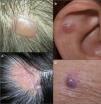
Case 1: A 68-year-old man attended the clinic because of an asymptomatic lesion that had appeared 6 months earlier in the right parietal region (Fig. 1A). The mildly erythematous nodule measured 1cm in diameter. The dermatoscopic image showed a homogeneous pattern of pale grey/bluish color, with whitish cotton wool-like structures and linear irregular vessels (Fig. 2A). Histopathology showed a cystic structure lined by an epithelium formed of a double layer of cuboidal cells with no cytologic atypia and areas of decapitation secretion, consistent with diagnosis of apocrine hidrocystoma (Fig. 3A).

Figure 1.

Clinical images. A, Nodule in the right parietal region. B, Cystic lesion on the helix. C, Nodule on sebaceous nevus on the scalp. D, Nodule on the right arm.

(0.35MB).
Figure 2.

Dermatoscopic images. Homogeneous pattern of pale grey/bluish color, with whitish cotton -wool –-like structures and linear irregular vessels (A-D) and regions of brown-orange color (B and D).

(0.17MB).
Figure 3.

Histologic images (hematoxylin-eosin, original magnification ×20). A and B, Dermal cysts enveloped in a double layer of cuboidal cells. Well-defined tumor composed of cells with abundant eosinophilic cytoplasm and other cells of a more basaloid appearance. C, Cystic structures in the center of the lesion.

(0.23MB).

Case 2: A 62-year-old woman attended the clinic because of an asymptomatic lesion on the right helix that had been present for several years. The translucent cystic lesion measured 1cm in diameter (Fig. 1B). The dermatoscopic image showed a homogeneous pattern of pale grey/bluish color, with brown-orange regions to the left of the lesion, whitish cotton wool-like structures, and linear irregular vessels (Fig. 2C). Histology was consistent with apocrine hidrocystoma (Fig. 3B).

Case 3: A 30-year-old woman had a 1cm nodule on a sebaceous nevus in the interparietal region (Fig. 1C). The dermatoscopic image showed a homogeneous pattern of pale grey/bluish color, with whitish cotton wool-like structures, and linear irregular vessels (Fig. 2C). Histology confirmed the diagnosis of apocrine hidrocystoma.

Case 4: An 81-year-old woman attended the clinic because of an asymptomatic lesion on the right arm. Clinical examination revealed a well-defined, erythematous nodule measuring 1.5cm in diameter (Fig. 1D). The dermatoscopic image showed a homogeneous pattern of pale grey/bluish color, with brown-orange areas, whitish cotton wool-like structures, and linear irregular vessels peripherally (Fig. 2D). Histologically, the lesion was a nonencapsulated dermal tumor comprised of cells with no atypia and abundant eosinophilic cytoplasm and cells whose nuclei had a more basaloid and hyperchromatic appearance. Foci of clear cells could be seen. The lesion showed cystic formations with mucin in the lumen, as well as a sclerotic and vascularized stroma (Fig. 3C). The above was compatible with a diagnosis of solid-cystic apocrine hidradenoma.

Apocrine hidrocystomas are benign adnexal cystic tumors of unknown etiology. Possible causes include sweat duct occlusion or adenomatous cystic proliferation of apocrine glands (apocrine cystadenoma). Histologically, they usually have multiple cystic spaces, with papillary protrusions and a wall composed of an inner layer of cuboidal cells and an outer layer of myoepithelial cells. These lesions are usually surrounded by a fibrous stroma with small-diameter vessels inside.2,3

Apocrine hidradenoma is a benign adnexal tumor of uncertain histogenesis. It is a multilobed, nonencapsulated, well-defined neoplasm comprised mainly of neoplastic epithelial cells and small ductal lumens. There are several histologic patterns: nodular hidradenoma, solid-cystic hidradenoma, and clear-cell hidradenoma.1,4,5

Correct diagnosis of hidrocystoma and hidradenoma is difficult solely on the clinical presentation and subsequent histologic confirmation is required. The clinical differential diagnosis is very broad and includes conditions such as other adnexal tumors, epidermal cysts, basal cell carcinoma, and amelanocytic melanoma.1–5

Correia et al.6 described the dermatoscopic findings from 2 eccrine hidrocystomas, but they only reported the presence of a well-defined cystic lesion free of vessels. Although not mentioned in the text of the article, a homogeneous pale grey/bluish pattern could be observed in the photograph of one of the cases. There is also a dermatoscopic description of a case of clear-cell hidradenoma, in which the presence of purpuric areas and some linear and hairpin vessels are noted on the tumor surface.7 The purpuric areas correspond histologically to hemorrhage into the cystic spaces of the tumor. In addition, there is a description of a case of malignant nodular hidradenoma, in which polymorphic vascular structures were observed.8 The authors suggested that these can be seen in tumors of eccrine origin.

All of our cases (3 apocrine hidrocystomas and 1 solid-cystic hidradenoma) had a homogeneous pale grey/bluish pattern, whitish cotton wool-like structures, and linear vessels. In 2 cases, focal brown-orange areas were observed. No purpuric areas were observed in any of our cases. The greyish color arises probably because these tumors contain sialomucin, which causes a diffraction effect similar to that observed in Kaposi sarcoma. The whitish cotton wool-like structures are a reflection from the stroma or connective tissue, while the dilated vessels in the superficial dermis correspond to the peripheral linear vessels in the dermatoscopic image. The presence of clear cells laden with glycogen could be responsible for the brown-orange focal structures. In conclusion, we should suspect these tumors in the case of nodular lesions, particularly on the head. Dermatoscopy is a technique that can help in diagnosis, particularly in the absence of dermatoscopic findings suggestive of other clinically similar tumors such as basal cell carcinoma and amelanotic melanoma.

References
[1]
N.A. Obaidat, K.O. Alsaad, D. Ghazarian.
Skin adnexal neoplasms-part 2: an approach to tumours of cutaneous sweat glands.
J Clin Pathol, 60 (2007), pp. 145-159
[2]
N. Vashi, R. Mandal.
Giant multi-loculated apocrine hidrocystomas.
Dermatol Online J, 16 (2010), pp. 16
[3]
S. Anzai, M. Goto, S. Fujiwara, T. Da.
Apocrine hidrocystoma: a case report and analysis of 167 Japanese cases.
Int J Dermatol, 44 (2005), pp. 702-703
[4]
B. Nandeesh, T. Rajalakshmi.
A study of histopathologic spectrum of nodular hidradenoma.
Am J Dermatopathol, 34 (2012), pp. 461-470
[5]
B. Monteagudo, I.M. Antón-Badiola, M.J. Muñoz, C. Paredes-Suárez, M. Vázquez-Blanco.
Hidradenoma apocrino pigmentado.
Actas Dermosifiliogr, 96 (2005), pp. 50-51
[6]
O. Correia, A.F. Duarte, A.M. Barros, N. Rocha.
Multiple eccrine hidrocystomas: from a diagnosis to treatment: the role of dermatoscopy and botulinum toxin.
Dermatology, 219 (2009), pp. 77-79
[7]
Y. Yoshida, K. Nakashima, O. Yamamoto.
Dermoscopic features of clear cell hidradenoma.
Dermatology, 217 (2008), pp. 250-251
[8]
O. Bakar, U. Ince.
A rare tumor of the face: malignant nodular hidradenoma with dermoscopic features mimicking amelanotic melanoma.
J Cutan Med Sur, 15 (2011), pp. 167-171

Please cite this article as: Cieza-Díaz D, Avilés-Izquierdo J, Ciudad-Blanco C, Parra-Blanco V. Dermatoscopia de tumores apocrinos: presentación de 4 casos. Actas Dermosifiliogr. 2013;104:826–828.

Copyright © 2012. Elsevier España, S.L. and AEDV
Idiomas
Actas Dermo-Sifiliográficas
Opciones de artículo
Herramientas
es en

¿Es usted profesional sanitario apto para prescribir o dispensar medicamentos?

Are you a health professional able to prescribe or dispense drugs?