


A 65-year-old woman with a 13-year-history of primary biliary cirrhosis, systemic hypertension, dyslipidemia, and osteoporosis came to our clinic for evaluation of lesions that had developed 2 years earlier. The lesions had gradually appeared on the back of the neck and were asymptomatic.
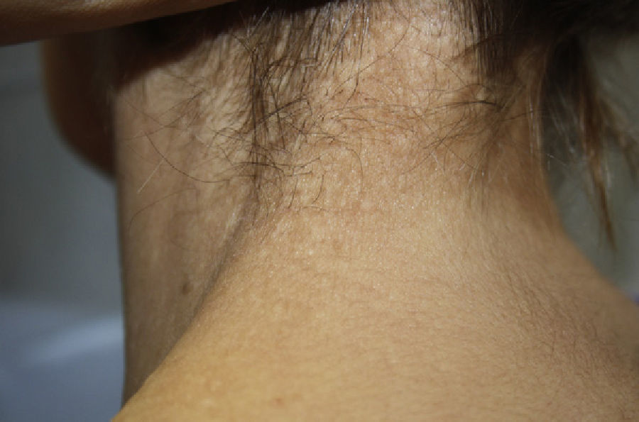
Physical ExaminationPhysical examination revealed multiple whitish papules measuring 3 mm in diameter and with a rubbery consistency; the follicular openings were spared and showed no mucinous material when pressed. The papules appeared in clusters on the back of the neck (Fig. 1), with occasional papules in the right axilla.
No other areas of the body were affected.
Additional TestsDermoscopy revealed well-delimited, homogenous, whitish papules with some dotted vessels at the periphery.
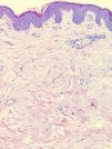
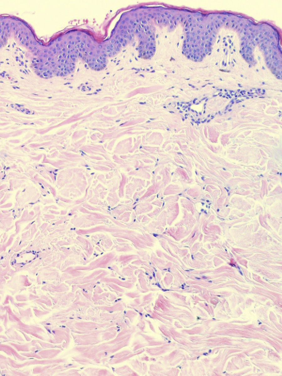
A 4-mm punch biopsy taken from a lesion on the neck showed slightly thickened collagen fibers in the mid dermis (Fig. 2). No calcium deposits were observed and staining of the elastic fibers revealed no abnormality.
What Is Your Diagnosis?
DiagnosisFibroelastolytic papulosis of the neck.
TreatmentGiven the benign nature of the lesions and the fact that they were asymptomatic, it was decided to adopt a conservative approach and no treatment was given.
CommentIn 1989 Shimizu et al.1 described white fibrous papulosis of the neck. Clinically this entity was characterized by isolated, asymptomatic whitish papules, mainly located on the back and side of the neck, although they were occasionally found on the shoulders and axillae. Histologically, white fibrous papulosis was classified under collagen disorders, given that the lesions were composed of circumscribed collagen bundles in the papillary and upper reticular dermis.
Rongioletti and Rebora2 later reported another very similar entity, which they named pseudoxanthoma elasticum-like papillary dermal elastolysis, in which they observed yellowish papules resembling those of white fibrous papulosis. In this case, however, the papules showed a tendency to coalesce, giving rise to cobblestone-like plaques suggestive of pseudoxanthoma elasticum (PXE). Histologically the lesions were mainly characterized by elastolysis, with no calcium deposits or fibrosis, and were categorized as elastic fiber abnormalities.
Balus et al.3 subsequently discovered common clinical and histologic findings when reviewing cases reported for both entities alongside 20 of their own. They concluded that there were 2 variants of a single condition that they named fibroelastolytic papulosis of the neck.
The exact cause of fibroelastolytic papulosis of the neck is unknown, although most authors have related it to the skin aging process. Recently cases have also been reported in younger patients with a history of inflammatory diseases in the same area. This suggests that factors other than aging, such as the inflammation of skin adnexa,4 can exert an influence on the subsequent appearance of fibrosis and elastolysis.
Diagnosis is based on clinical presentation, histologic findings, and, more recently, dermoscopy, which shows well-delimited, whitish papules with some dotted vessels at the periphery, as well as pigment that fails to form a true reticulated area.5
The case reported here is more compatible with what was originally known as white fibrous papulosis of the neck. This definitive diagnosis was established from the clinical presentation and dermoscopy, but particularly after the exclusion of other entities, such as PXE, on pathology study of the biopsy.
In conclusion, knowledge of fibroelastolytic papulosis of the neck is important as it bears a strong clinical resemblance to PXE. Unlike PXE, however, prognosis is good and not associated with heart or ophthalmological disease.
Please cite this article as: Echeverría-García B, et al. Pápulas en empedrado en el cuello. Actas Dermosifiliogr. 2013;104:247–8.


