


A 56-year-old man presented with multiple small asymptomatic whitish papules that had appeared 3 months earlier on both hands. The patient's past history included systemic hypertension on treatment with valsartan, untreated hyperuricemia, dyslipidemia, and moderate alcohol consumption with findings on magnetic resonance imaging suggestive of chronic pancreatitis.
Physical ExaminationPhysical examination showed numerous small (2-3mm) whitish papules on the palms and fingertips of both hands. The papules were hard and smooth and some of them were ulcerated (Fig. 1).
Additional Tests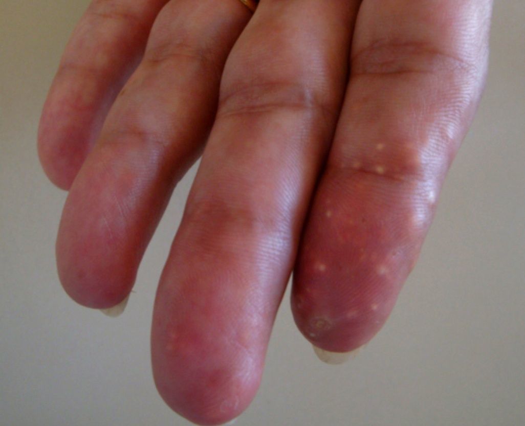
A biopsy was performed of 1 of the lesions. Microscopy revealed a large nodular deposit of amorphous material containing acicular clefts in the dermis, surrounded by pseudopalisading histiocytes and a marked foreign body giant cell reaction. In close proximity to the main lesion we observed multiple, small, coalescing nodular deposits comprised of similar material with a marked associated giant cell reaction. The epidermis showed no significant changes (Figs. 2 and 3).


What Is Your Diagnosis?
DiagnosisMilia-like cutaneous tophi
Clinical Course and TreatmentThe patient was referred to the rheumatology department and treated with allopurinol (100mg/d), resulting in a gradual improvement in the lesions.
CommentThe clinical features of gout are caused by deposits of monosodium urate crystals in the tissues. The main clinical manifestations of gout are gouty arthritis, the accumulation of crystals in the connective tissues (tophi), uric acid nephrolithiasis, and renal failure.1 Tophi appear after a long period of hyperuricemia. The prevalence of this condition has been reduced by effective therapies and is now limited to patients who fail to comply with treatment and those in whom the diagnosis of gout is delayed.2 Rarely, tophi may be the first sign of hyperuricemia.3 Tophi appear as whitish-yellowish dermal papules or subcutaneous nodules that are firm to the touch. The borders may be smooth or lobulated. They appear most frequently on the skin covering the joints and on the helix of the ear. Presentation as numerous small papules appearing rapidly on the hands is very rare. Historically, this condition has been referred to as intradermal tophi or pustular gouty tophi. The term milia-like tophi has recently been proposed for this entity based on the similarity between these lesions and millet seeds as well as the wider distribution of the lesions involving not only the fingertips, which has been previously described, but also the palms.4 The main histologic feature of gouty tophi is the presence deposits of an amorphous material within the dermis and subcutaneous cellular tissue. These deposits contain acicular clefts, caused by the dissolution of urate crystals, and are surrounded by an infiltrate comprised of histiocytes, in the form of multinucleated giant cells, and lymphocytes, sometimes with the presence of a fibrous capsule. The epidermis may be intact or ulcerated. During standard processing and fixing techniques most of the urate crystals are dissolved and fixation in ethanol or freezing of the tissue is therefore necessary to identify the crystals.5
When observed under a polarized light filter, the acicular crystals exhibit negative birefringence. The clinical differential diagnosis of gouty tophi should include xanthomas, rheumatoid nodules, and calcinosis cutis. The prolonged use of medications that reduce uric acid levels such as allopurinol can successfully treat newer, smaller lesions,6 while larger lesions may be removed surgically.
Conflict of InterestThe authors declare that they have no conflicts of interest.
Please cite this article as: Bernat García J, et al. Pápulas blanquecinas en las manos. Actas Dermosifiliogr. 2013;104:349–50.


