Journal Information
Vol. 103. Issue 6.
Pages 488-501 (July - August 2012)
Visits
17730
Vol. 103. Issue 6.
Pages 488-501 (July - August 2012)
Practical Dermatology
Full text access
Photodynamic Therapy vs Imiquimod
Terapia fotodinámica versus imiquimod
Visits
17730
C. Serra-Guillén
Corresponding author
cserraguillen@gmail.com

Corresponding author.
, E. Nagore, C. Guillén
Servicio de Dermatología, Instituto Valenciano de Oncología, Valencia, Spain
This item has received
Article information
Abstract
Full Text
Bibliography
Download PDF
Statistics
Figures (8)
Show moreShow less
Tables (3)
Table 1. Studies of Patients With Bowen Disease Treated With Photodynamic Therapy or Imiquimod.
Table 2. Studies of Superficial Basal Cell Carcinoma Treated With Photodynamic Therapy or Imiquimod.
Table 3. Studies of Patients with Nodular Basal Cell Carcinoma Treated With Photodynamic Therapy and Imiquimod.
Show moreShow less
Abstract

Photodynamic therapy and imiquimod are highly regarded treatments dermatologists frequently prescribe for actinic keratoses, basal cell carcinoma, and Bowen disease. The scarcity of evidence from comparative trials prevents us from drawing well-founded conclusions about the efficacy, tolerance, and adverse effects of these therapeutic options or to recommend one over the other in any particular type of lesion or patient. On the other hand, in certain conditions (eg, actinic chelitis, immunosuppression, and basal cell carcinoma affecting the eyelids), there is evidence to support the use of photodynamic therapy or imiquimod even though they might initially seem contraindicated. We critically review and compare the use of these 2 treatments in order to suggest which is more appropriate in specific cases.

Keywords:
Photodynamic therapy
Imiquimod
Actinic keratosis
Basal cell carcinoma
Bowen disease
Treatment
Resumen

La terapia fotodinámica (TFD) y el imiquimod son dos excelentes tratamientos utilizados frecuentemente en Dermatología para las queratosis actínicas, el carcinoma basocelular (CBC) o la enfermedad de Bowen. No existen suficientes estudios comparativos entre ellos para poder extraer buenas conclusiones sobre su eficacia, su tolerancia o sus efectos secundarios y para poder situar a un tratamiento por encima del otro en un tipo de lesión o paciente en concreto. Por otra parte, existen situaciones o indicaciones particulares como la queilitis actínica, los pacientes inmunodeprimidos o los CBC localizados en párpados donde estos dos tratamientos pueden considerarse inicialmente contraindicados; sin embargo, existe suficiente evidencia para poder utilizarlos.

Vamos a realizar una revisión de la TFD y el imiquimod, bajo un punto de vista crítico y comparativo entre ellos, para poder ayudar a responder a la cuestión de qué tratamiento es más recomendable en un paciente determinado.

Palabras clave:
Terapia fotodinámica
Imiquimod
Queratosis actínica
Carcinoma basocelular, Enfermedad de Bowen
Tratamiento
Full Text
Introduction

Photodynamic therapy (PDT) and imiquimod are 2 highly regarded treatments with very similar indications in dermatology. While many studies have compared one or other of these therapies with other treatment options—for instance, cryotherapy,1–3 fluorouracil,3,4 or surgery5,6—very few have compared PTD with imiquimod.7,8 It is not unusual in routine clinical practice for clinicians to have difficulty deciding which of these therapies is more appropriate in a specific case since both are routinely used to treat actinic keratoses (AKs), basal cell carcinoma (BCC), and Bowen disease (BD). Several small case series and anecdotal case reports have described the use of PDT and/or imiquimod in other skin cancers, including lentigo maligna,9 mycosis fungoides,10 and Paget disease,11 as well as in benign skin diseases.12 However, the lack of scientific evidence for their use in these settings and the existence of more appropriate treatment options make it unlikely that the list of approved indications for PDT or imiquimod will be modified, a step that would allow these treatments to be used more often and with greater safety.

The initial step in PDT is the application of a photosensitizing agent, either aminolevulinic acid (ALA) or methyl-aminolevulinic acid (MAL), to the lesion to be treated. This is then incubated under an occlusive dressing for at least 3hours and subsequently illuminated, usually with a light having a wavelength in the 570 to 670nm range. PDT with MAL (Metvix cream, Galderma SA) is approved for AKs, superficial and nodular BCC, and BD according to the Summary of Product Characteristics (SPC). The therapeutic indication in the case of AKs is nonhyperkeratotic lesions on the face or scalp. The recommended regimen is a single session, which can be repeated after a 3-month interval if needed. In the treatment of BD and superficial and/or nodular BCC, 2 PDT sessions separated by an interval of at least 1 week are recommended.

Imiquimod is sold in the form of a 5% cream (Aldara cream, MEDA AB), which is applied by the patient. It is approved for the treatment of condylomata acuminata, nonhyperkeratotic, nonhypertrophic AKs on the face or scalp in immunocompetent patients, and small superficial BCCs (the SPC does not specify the size). The posology specified in the case of AKs is once-daily application 3 times a week for 4 weeks. If no response is observed on follow-up, the 4-week treatment cycle may be repeated. In the treatment of superficial BCCs, imiquimod should be applied 5 times a week for 6 weeks.

We review the use of PDT and imiquimod in the approved indications. We also discuss their use in other settings in which they might initially appear to be contraindicated, such as actinic cheilitis, tumors in transplant recipients, and BCCs on the eyelids. The safety and efficacy of these treatments in these settings have, however, been demonstrated, making it possible to consider their use in selected cases. The benefits and drawbacks of the 2 treatments are analyzed to provide a basis for decisions regarding the most appropriate choice of treatment in a given situation.

Actinic Keratoses

AKs are the precancerous lesions most frequently encountered by dermatologists. They often appear in the context of field cancerization. This term is used to describe areas of chronically exposed skin characterized by the presence of AKs and histologic changes. In addition to dysplastic keratinocytes, the histologic features include molecular changes such as p53 mutations that predispose patients to squamous cell carcinomas but are not manifest in the form of clinically apparent lesions.13,14 PDT and imiquimod are the first-line treatments in these patients as they can be used to treat large areas of skin and subclinical lesions at the same time. The cosmetic results obtained with both treatments are also excellent.3,15,16 However, there are some differences that may influence the decision to use one treatment or the other.

The use of PDT in the treatment of AKs has been studied by numerous authors, but it is difficult to accurately assess overall treatment efficacy because of differences in the methodologies used in each study. Cure rates after 1 or 2 treatment sessions using MAL as the photosensitizing agent were 69% to 91% (Fig. 1).15,17–20 The adverse effect most often reported with PDT is pain during the illumination phase of treatment. Pain tends to be more severe in patients with skin phototypes I and II and when the lesions treated are on the head.21 However, as in any other dermatologic procedure, there are a variety of effective methods for controlling or reducing pain, including cold air and nerve block anesthesia.22 Thus, the pain caused by PDT should not be a reason for avoiding the use of this treatment modality. Interestingly, despite the widespread impression that its tolerance can be problematic, PDT is the highest-rated treatment for AKs in terms of patient satisfaction in all the studies that have evaluated this parameter.1,8,15,19,23 This high degree of patient satisfaction is probably due to the fact that a PDT session takes up very little time and evidence of improvement can be observed within a few weeks. Likewise, PDT is not a treatment patients have to apply themselves and is therefore a modality usually not associated with doubts on the part of the patient or the occurrence of adverse effects at home which they are unable to cope with.

Figure 1.

Multiple actinic keratoses treated with methyl aminolevulinate photodynamic therapy. A, Prior to treatment. B, Illumination. C, Results after 1 photodynamic therapy session.

(0.22MB).

It is difficult to evaluate the long-term recurrence rate in patients with AKs treated with PDT because very few of the studies in the literature have a follow-up period longer than 12 months.24–26 Rates of up to 24% at 1 year have been reported,27 but recurrence is lower with fractionated illumination28,29 and nonhyperkeratotic lesions.30

In transplant recipients, PDT has proved very useful in curing AKs31,32 and in preventing new lesions.33,34 Moreover, some studies have shown cyclic application of PDT to reduce the risk of squamous cell carcinoma in these patients.35 However, other authors have demonstrated that PDT does not reduce the incidence of squamous cell carcinomas in transplant recipients with actinic damage.36

The safety and efficacy of PDT in the treatment of actinic cheilitis (Fig. 2) have been demonstrated in many studies. Good response and good tolerance have been reported following 2 or 3 PDT sessions with either MAL37,38 or ALA,39,40 leading some authors to consider PDT the treatment of choice in actinic chelitis.41 However, in the studies of PDT in which histologic confirmation of response was obtained, lesions persisted in 20% to 53% of patients37,42 and histologic changes compatible with actinic cheilitis were found after 18 months of follow up in 34.6% of the patients treated.40 Based on the evidence cited above, PDT as a treatment for actinic cheilitis has a B strength recommendation rating and should be considered a second-line treatment.43

Figure 2.

Actinic cheilitis treated with photodynamic therapy using methyl aminolevulinate. A, Prior to treatment. B, Illumination. C, Results after 1 photodynamic therapy session.

(0.17MB).

It is likewise difficult to determine the exact cure rate achieved with imiquimod in the treatment of AKs. Most studies report complete or partial clinical response rates, while others report histologic response. Furthermore, treatment regimens vary greatly from one study to another. Three meta-analyses have found imiquimod to be a very effective treatment for AKs, achieving complete response (100% of the lesions resolved after treatment) in more than 70% of patients (Fig. 3).44–46 One of the most important characteristics of imiquimod treatment is the high negative predictive value of the assessment of the clinical response. The methodology used in many studies of imiquimod appears to be more rigorous than that used in studies of PDT, and histologic confirmation of treatment response is more often included. The probability that AKs that are clinically resolved with imiquimod are also histologically resolved ranges from 86% to 100%.3,47–49 It is interesting to note that studies of imiquimod in AK have longer follow-up periods than studies of PDT in this setting. More imiquimod studies have assessed clinical response 1 year after completion of treatment, when most authors report a recurrence rate close to 10%,3,48,50 although Jorizzo et al.51 reported a recurrence rate of 39% at 1 year. The main adverse effect associated with imiquimod is a local inflammatory reaction that occurs during treatment. This reaction, which is necessary and even predictive of a good reponse,44 appears within a few days of treatment initiation and takes the form of erythema, inflammation, swelling, and even crusting (Fig. 4). In addition, some patients develop flu-like symptoms, such as general malaise, fever, and muscle weakness. These adverse effects appear to be the main reason why some clinicians are reluctant to prescribe imiquimod to patients with AKs. The treatment cycle lasts for 4 weeks, and the local reaction may last as long as 2 months. Thus, patients and their families need to be properly informed so that they will understand the process involved. This precaution will also prevent unnecessary phone calls, unscheduled visits to the physician, and even visits to the emergency department, where symptoms are often misinterpreted and treatment discontinued. This adverse reaction, which has been widely studied in protocols and studies, has been reported in almost 100% of patients in the majority of publications.44,46–48 Paradoxically, however, most authors have concluded that imiquimod is well tolerated in the treatment of AKs.48,52,53 In the only 2 studies comparing PDT and imiquimod that have evaluated patient preference7 and satisfaction,8 more patients favored PDT. Researchers investigating ways to reduce the local adverse effects of imiquimod while maintaining its efficacy have evaluated creams with reduced concentrations of the active ingredient—2.5% or 3.75%—with promising results.54,55 However, these formulations are not currently available in Europe.

Figure 3.

Multiple actinic keratoses treated with imiquimod. A, Prior to treatment. B, 3 months after completion of treatment. C, Prior to treatment. D, 3 months after completion of treatment.

(0.3MB).
Figure 4.

A-C. Local reaction at week 4 of treatment with imiquimod for multiple actinic keratoses.

(0.24MB).

According to the SPC, the use of imiquimod is contraindicated in transplant recipients because, in theory, the drug's mechanism of action relies on the patient having an adequate immune system. Nevertheless, some studies of the safety and efficacy of imiquimod in transplant recipients have reported more than acceptable results in the treatment of AKs,56,57 and even in preventing the development of squamous cell carcinoma.58 Moreover, there is scant evidence in the literature to suggest that imiquimod may produce local or systemic immunologic changes in the patient, skin tumors in the treatment area,59 or autoimmune disorders.60 Most studies and reviews of the literature accept and recommend the use of imiquimod in immunosuppressed patients.57,61

Imiquimod may also be considered in the treatment of actinic cheilitis, although there are fewer studies on its use in this setting than in the case of PDT. According to the 2 largest series of patients studied (1562 and 563), imiquimod appears to be clinically and histologically effective when applied for at least 4 weeks. However, the use of imiquimod on the lips is greatly limited by the visible local reaction that almost always accompanies treatment and the possibility that the patient may develop oral aphthous ulcers64 or experience an outbreak of herpes simplex43 during treatment.

In conclusion, both PDT and imiquimod are the best treatments available for patients with multiple AKs or field cancerization. Their efficacy is similar although there are certain differences that should be taken into account when deciding which option is most suitable in a particular patient or situation. PDT is superior in terms of tolerance and patient satisfaction, but imiquimod appears to be associated with lower recurrence. While both PDT and imiquimod are effective in the treatment of AKs in immunosuppressed transplant recipients, PDT would appear to be a slightly better choice because of its mechanism of action and the larger number of studies supporting its efficacy in curing AKs and in preventing both new lesions and the development of squamous cell carcinomas. In the case of actinic cheilitis, imiquimod is not recommended because of the associated adverse effects; PDT, by contrast is a recommendable alternative in many patients but surgical treatment remains the method of choice in this setting.
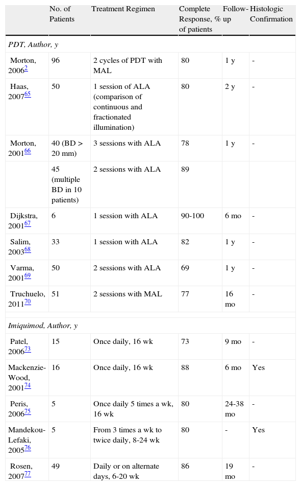
Bowen Disease

The efficacy of PDT in the treatment of BD is between 69% and 100% (Fig. 5).2,65–70 Most of the studies reviewed involved 2 treatment sessions and used ALA as the photosensitizer. In the largest multicenter study in the literature, Morton et al.2 reported an 80% complete response rate at 12 months following 2 sessions of PDT with MAL. Many studies report excellent cosmetic results in most cases.2,70 Given that these lesions are usually small (only a few centimeters) and the fact that they are not located on the head, tolerance of treatment is generally very good.21,71 The British Association of Dermatologists (BAD) guidelines for the management of BD has assigned PDT the highest rating of all the treatment options reviewed (quality of evidence rating I and strength of recommendation A).72

Figure 5.

Bowen disease treated with photodynamic therapy using methyl aminolevulinate. A, Prior to treatment. B, Illumination. C, Results after 3 photodynamic therapy sessions.

(0.16MB).

Five studies of patients with BD treated with imiquimod have been published (Fig. 6).73–77 Overall, treatment lasted from 9 to 16 weeks and complete response was obtained in 73% to 88% of the patients studied, in some cases with histologic confirmation; the longest follow-up period was 19 months. Tolerance was generally acceptable, although a few patients discontinued treatment because of the local reaction. The BAD guidelines assigned treatment with imiquimod a quality of evidence rating of I and a strength of recommendation rating of B.72

Figure 6.

Bowen disease treated with imiquimod. A, Prior to treatment. B, Local reaction after 3 weeks of treatment. C. Result 3 months after completion of treatment.

(0.16MB).

Table 1 lists the most important studies of PDT and imiquimod in the treatment of BD.

Table 1.

Studies of Patients With Bowen Disease Treated With Photodynamic Therapy or Imiquimod.

  No. of Patients  Treatment Regimen  Complete Response, % of patients  Follow-up  Histologic Confirmation 
PDT, Author, y
Morton, 20062  96  2 cycles of PDT with MAL  80  1 y 
Haas, 200765  50  1 session of ALA (comparison of continuous and fractionated illumination)  80  2 y 
Morton, 200166  40 (BD>20mm)  3 sessions with ALA  78  1 y 
  45 (multiple BD in 10 patients)  2 sessions with ALA  89     
Dijkstra, 200167  1 session with ALA  90-100  6 mo 
Salim, 200368  33  1 session with ALA  82  1 y 
Varma, 200169  50  2 sessions with ALA  69  1 y 
Truchuelo, 201170  51  2 sessions with MAL  77  16 mo 
Imiquimod, Author, y
Patel, 200673  15  Once daily, 16 wk  73  9 mo 
Mackenzie-Wood, 200174  16  Once daily, 16 wk  88  6 mo  Yes 
Peris, 200675  Once daily 5 times a wk, 16 wk  80  24-38 mo 
Mandekou-Lefaki, 200576  From 3 times a wk to twice daily, 8-24 wk  80  Yes 
Rosen, 200777  49  Daily or on alternate days, 6-20 wk  86  19 mo 

aComplete response rates refer to the result at the end of the follow-up period specified.

Abbreviations: ALA, aminolevulinic acid; BD, Bowen disease; MAL methyl aminolevulinate.

In the treatment of BD, PDT is generally preferred over imiquimod for a number of reasons, as indicated in the BAD guidelines for this disease.72 PDT is superior to imiquimod in this setting, first because, strictly speaking, it is approved for this indication while imiquimod is not. Furthermore, more patients have been studied in the case of PDT and the rate of complete response to treatment is slightly higher. Nonetheless, treatment with both PDT and imiquimod should be considered before surgical intervention in patients with BD. It would seem unreasonable to propose surgery when 2 conservative choice of treatment should take into account the site of the lesion (for example, in the case of BD affecting the perianal region, the ears, or other areas where illumination may be complicated), the availability of the patient to attend PDT sessions or, in the case of imiquimod, the patient's ability to properly apply treatment.
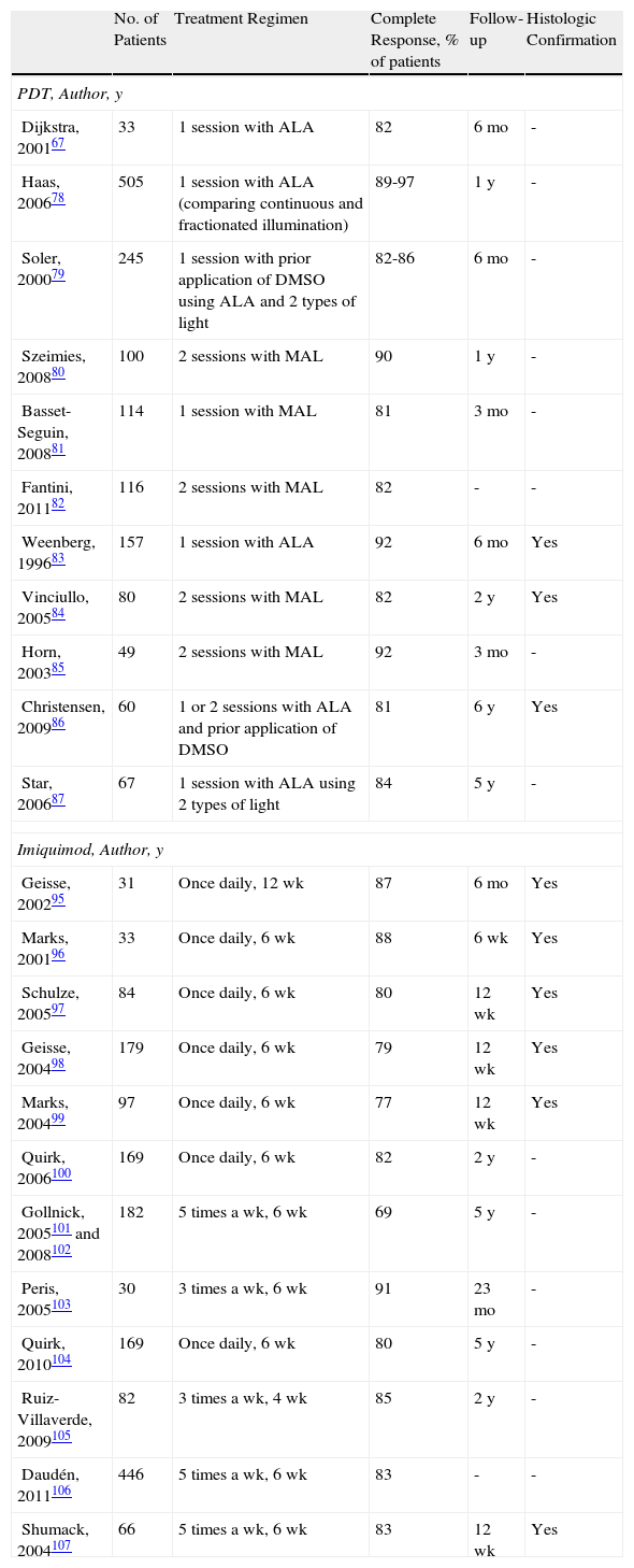
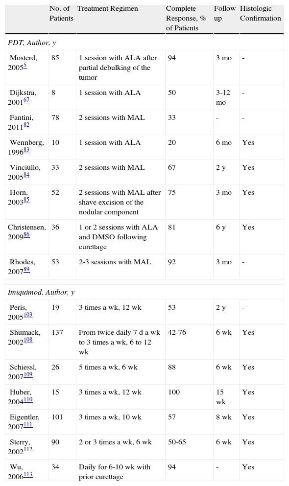
Basal Cell Carcinoma

A review of studies of PDT in the treatment of BCC also reveals methodological differences between studies. Some authors have used ALA and others MAL. The light sources and the methods used to apply the light also differ, as do methods for assessing response. However, there is consensus on the recommended regimen of 2 sessions of PDT with ALA or MAL separated by an interval of at least 1 week. It is also generally accepted that superficial BCC responds very well to 2 sessions of PDT using ALA or MAL. Response rates of 81% to 97% were reported for these treatments in the largest case series.67,78–87 On the basis of this evidence, the BAD guidelines recommend PDT as the first-line choice for the treatment of BCC (quality of evidence I and strength of recommendation A).88 However, the response rates reported for nodular BCC are lower than for superficial BCC, with complete response in between 20% to 94% of patients depending on the study.5,67,82–86,89 Furthermore, it could be said that these figures do not entirely reflect the real efficacy of PDT because—in the studies reporting the best results—lesions were pretreated with various procedures, such as the application of dimethyl sulfoxide,86 debulking,5 curettage,86 or shaving of the tumor.85 Furthermore, a study of recurrence during follow-up periods ranging from 1 to 6 years reveals a recurrence rate for superficial BCC of between 3% and 22%78,80,84–86 and a somewhat higher rate (14%-33%) for nodular BCC.5,84–86,89 Consequently, PDT is considered to be a second-line treatment for nodular BCC, (quality of evidence I, strength of recommendation B) with surgery as the first-line treatment (quality of evidence I, strength of recommendation A).88

Another interesting conclusion that can be drawn from the findings of the studies on PDT in BCC is that fractionation of the illumination phase into 2 exposures separated by a dark interval of varying length greatly improves response; this conclusion has relatively broad acceptance.5,29,78,87

Tables 2 and 3 list the most important studies of PDT and imiquimod in the treatment of superficial and nodular BCC.

Table 2.

Studies of Superficial Basal Cell Carcinoma Treated With Photodynamic Therapy or Imiquimod.

  No. of Patients  Treatment Regimen  Complete Response, % of patients  Follow-up  Histologic Confirmation 
PDT, Author, y
Dijkstra, 200167  33  1 session with ALA  82  6 mo 
Haas, 200678  505  1 session with ALA (comparing continuous and fractionated illumination)  89-97  1 y 
Soler, 200079  245  1 session with prior application of DMSO using ALA and 2 types of light  82-86  6 mo 
Szeimies, 200880  100  2 sessions with MAL  90  1 y 
Basset-Seguin, 200881  114  1 session with MAL  81  3 mo 
Fantini, 201182  116  2 sessions with MAL  82 
Weenberg, 199683  157  1 session with ALA  92  6 mo  Yes 
Vinciullo, 200584  80  2 sessions with MAL  82  2 y  Yes 
Horn, 200385  49  2 sessions with MAL  92  3 mo 
Christensen, 200986  60  1 or 2 sessions with ALA and prior application of DMSO  81  6 y  Yes 
Star, 200687  67  1 session with ALA using 2 types of light  84  5 y 
Imiquimod, Author, y
Geisse, 200295  31  Once daily, 12 wk  87  6 mo  Yes 
Marks, 200196  33  Once daily, 6 wk  88  6 wk  Yes 
Schulze, 200597  84  Once daily, 6 wk  80  12 wk  Yes 
Geisse, 200498  179  Once daily, 6 wk  79  12 wk  Yes 
Marks, 200499  97  Once daily, 6 wk  77  12 wk  Yes 
Quirk, 2006100  169  Once daily, 6 wk  82  2 y 
Gollnick, 2005101 and 2008102  182  5 times a wk, 6 wk  69  5 y 
Peris, 2005103  30  3 times a wk, 6 wk  91  23 mo 
Quirk, 2010104  169  Once daily, 6 wk  80  5 y 
Ruiz-Villaverde, 2009105  82  3 times a wk, 4 wk  85  2 y 
Daudén, 2011106  446  5 times a wk, 6 wk  83 
Shumack, 2004107  66  5 times a wk, 6 wk  83  12 wk  Yes 

aComplete response rates refer to the result at the end of the follow-up period specified.

Abbreviations: ALA, aminolevulinic acid; DMSO, dimethyl sulfoxide; MAL, methyl aminolevulinate.

Table 3.

Studies of Patients with Nodular Basal Cell Carcinoma Treated With Photodynamic Therapy and Imiquimod.

  No. of Patients  Treatment Regimen  Complete Response, % of Patients  Follow-up  Histologic Confirmation 
PDT, Author, y
Mosterd, 20055  85  1 session with ALA after partial debulking of the tumor  94  3 mo 
Dijkstra, 200167  1 session with ALA  50  3-12 mo 
Fantini, 201182  78  2 sessions with MAL  33 
Wennberg, 199683  10  1 session with ALA  20  6 mo  Yes 
Vinciullo, 200584  33  2 sessions with MAL  67  2 y  Yes 
Horn, 200385  52  2 sessions with MAL after shave excision of the nodular component  75  3 mo  Yes 
Christensen, 200986  36  1 or 2 sessions with ALA and DMSO following curettage  81  6 y  Yes 
Rhodes, 200789  53  2-3 sessions with MAL  92  3 mo 
Imiquimod, Author, y
Peris, 2005103  19  3 times a wk, 12 wk  53  2 y 
Shumack, 2002108  137  From twice daily 7 d a wk to 3 times a wk, 6 to 12 wk  42-76  6 wk  Yes 
Schiessl, 2007109  26  5 times a wk, 6 wk  88  6 wk  Yes 
Huber, 2004110  15  3 times a wk, 12 wk  100  15 wk  Yes 
Eigentler, 2007111  101  3 times a wk, 10 wk  57  8 wk  Yes 
Sterry, 2002112  90  2 or 3 times a wk, 6 wk  50-65  6 wk  Yes 
Wu, 2006113  34  Daily for 6-10 wk with prior curettage  94  Yes 

aComplete response rates refer to the result at the end of the follow-up period specified.

Abbreviations: ALA, aminolevulinic acid; DMSO, dimethyl sulfoxide; MAL, methyl aminolevulinate.

The use of PDT in the periocular area, and even to treat tumors located on the eyelids, might initially appear to be absolutely contraindicated because of the possibility of patient discomfort and the technical difficulties involved. However, based on the available evidence90–93 and our own experience, our opinion is that PDT can be used in selected patients with superficial and nodular BCC in these areas when surgery is contraindicated, but only if the eye is shielded with a protective lens before the photosensitizer is applied (Fig. 7). Tolerance in such cases is generally good, and PDT can be used as a neoadjuvant, palliative, or even curative therapy.

Figure 7.

Ulcerated and nodular basal cell carcinoma on the right lower eyelid treated with photodynamic therapy using methyl aminolevulinate. A, Prior to treatment. B, Illumination. C, Results after 3 photodynamic therapy sessions.

(0.2MB).

The treatment of superficial BCC with imiquimod has been widely studied. A review of the literature indicates that the methodology used in these studies appears to have been more rigorous and uniform than that used in studies of PDT. In superficial BCC, there are also slightly more studies on the use of imiquimod than of PDT.94 In the largest studies, the rate of complete response in cases of superficial BCC treated with imiquimod ranged from 69% to 91%95–107 (quality of evidence I and strength of recommendation A).88 In most of these studies, response to treatment was confirmed by histologic evidence95–99,107 or analyzed in a follow-up period of up to 5 years,101,102,104 enhancing the reliability of the results regarding the efficacy of imiquimod in the treatment of superficial BCC. However, as occurs in the case of AKs, treatment of BCC with imiquimod causes a local reaction that may last for up to 2 months; this adverse effect often prevents the patient from correctly completing treatment and thus limits the use of this therapy in BCC.

Although imiquimod is not approved for the treatment of nodular BCC, there is nonetheless sufficient evidence and clinical experience to support its use. In the largest studies reviewed, treatment with imiquimod resulted in complete response in between 42% and 100% of cases of nodular BCC.103,108–113 As in the case of PDT, prior curettage has been proposed as a way of increasing the efficacy of imiquimod in this setting.113,114

As is the case with PDT, the use of imiquimod to treat BCCs located in a periocular site or on the eyelids might at first appear to be absolutely contraindicated. However, many authors have reported results that support the use of imiquimod in selected cases (Fig. 8).115–119 The studies of García-Martín et al.118 and Cannon et al.119 are of particular interest. García-Martín et al. reported complete clinical and histologic response in all 15 cases of periocular BCC treated with imiquimod, with excellent cosmetic results. Cannon et al. analyzed the adverse effects in 47 patients treated with imiquimod for periocular tumors. The most common effect was conjunctivitis of medium severity (11 patients). All adverse effects resolved once treatment was stopped, and there were no residual adverse effects.

Figure 8.

Superficial basal cell carcinoma on the right lower eyelid treated with imiquimod. A, Prior to treatment. B, Local reaction and mild conjunctivitis after 3 weeks of treatment. C, Local reaction and mild conjunctivitis after 6 weeks of treatment. D, Clinical response and resolution of conjunctivitis 1 month after completion of treatment with imiquimod.

(0.4MB).

Both PDT and imiquimod are less costly options than surgical excision in the treatment of BCC,6,120,121 and PDT is more economical than imiquimod.6

In conclusion, both PDT and imiquimod should be considered first-line treatments for superficial BCC in terms of efficacy. Surgery should only be used when the disease proves refractory to these treatments or when the site of the tumor is a contraindication for these treatments, a specimen is required for histologic examination, or the patient prefers surgery. Several factors come into play in the decision whether to choose PDT or imiquimod to treat superficial BCCs. Taking into account the greater ease of treatment, patient preferences, and the cosmetic results obtained, PDT provides greater benefits and should, therefore, generally be preferred to treatment with imiquimod in this setting.25,88,122 It should be noted that neither PDT nor imiquimod offers greater efficacy than surgery in the treatment of nodular BCC. When surgery is contraindicated in nodular BCC, PDT is rated as a better choice of treatment than imiquimod in the BAD guidelines for the management of BCC and in the SPCs of both products. Finally, in selected patients, both PDT and imiquimod can be attractive alternatives for the treatment of nodular and superficial BCCs on the eyelids and in the periocular area.

Conflicts of Interest

The authors declare that they have no conflicts of interest.

References
[1]
R. Kaufmann, L. Spelman, W. Weightman, J. Reifenberger, R.M. Szeimies, E. Verhaeghe, et al.
Multicentre intraindividual randomized trial of topical methyl aminolaevulinate-photodynamic therapy vs cryotherapy for multiple actinic keratoses on the extremities.
Br J Dermatol, 158 (2008), pp. 994-999
[2]
C. Morton, M. Horn, J. Leman, B. Tack, C. Bedane, M. Tjioe, et al.
Comparison of topical methyl aminolevulinate photodynamic therapy with cryotherapy or Fluorouracil for treatment of squamous cell carcinoma in situ: Results of a multicenter randomized trial.
Arch Dermatol, 142 (2006), pp. 729-735
[3]
N. Krawtchenko, J. Roewert-Huber, M. Ulrich, I. Mann, W. Sterry, E. Stockfleth.
A randomised study of topical 5% imiquimod vs. topical 5-fluorouracil vs. cryosurgery in immunocompetent patients with actinic keratoses: a comparison of clinical and histological outcomes including 1-year follow-up.
Br J Dermatol, 157 (2007), pp. 34-40
[4]
H.A. Kurwa, S.A. Yong-Gee, P.T. Seed, A.C. Markey, R.J. Barlow.
A randomized paired comparison of photodynamic therapy and topical 5-fluorouracil in the treatment of actinic keratoses.
J Am Acad Dermatol, 41 (1999), pp. 414-418
[5]
K. Mosterd, M.R. Thissen, P. Nelemans, N.W. Kelleners-Smeets, R.L. Janssen, K.G. Broekhof, et al.
Fractionated 5-aminolaevulinic acid-photodynamic therapy vs surgical excision in the treatment of nodular basal cell carcinoma: results of a randomized controlled trial.
Br J Dermatol, 159 (2008), pp. 864-870
[6]
M. Aguilar, M. de Troya, L. Martin, N. Benítez, M. González.
A cost analysis of photodynamic therapy with methyl aminolevulinate and imiquimod compared with conventional surgery for the treatment of superficial basal cell carcinoma and Bowen's disease of the lower extremities.
J Eur Acad Dermatol Venereol, 24 (2010), pp. 1431-1436
[7]
E. Sotiriou, Z. Apalla, F. Maliamani, N. Zaparas, D. Panagiotidou, D. Ioannides.
Intraindividual, right-left comparison of topical 5-aminolevulinic acid photodynamic therapy vs 5% imiquimod cream for actinic keratoses on the upper extremities.
J Eur Acad Dermatol Venereol, 23 (2009), pp. 1061-1065
[8]
C. Serra-Guillén, E. Nagore, L. Hueso, B. Llombart, C. Requena, O. Sanmartín, et al.
A randomized comparative study of tolerance and satisfaction in the treatment of actinic keratosis of the face and scalp between 5% imiquimod cream and photodynamic therapy with methyl aminolaevulinate.
Br J Dermatol, 164 (2011), pp. 429-433
[9]
A.M. Powell, A.M. Robson, R. Russell-Jones, R.J. Barlow.
Imiquimod and lentigo maligna: a search for prognostic features in a clinicopathological study with long-term follow-up.
Br J Dermatol, 160 (2009), pp. 994-998
[10]
M. Fernández-Guarino, A. Harto, B. Pérez-García, C. Montull, E. De Las Heras, P. Jaen.
Plaque-phase mycosis fungoides treated with photodynamic therapy: results from 12 patients.
Actas Dermosifiliogr, 101 (2010), pp. 785-791
[11]
E. Sendagorta, P. Herranz, M. Feito, P. Ramírez, U. Floristan, R. Feltes, et al.
Successful treatment of three cases of primary extramammary Paget‘s disease of the vulva with Imiquimod--proposal of a therapeutic schedule.
J Eur Acad Dermatol Venereol, 24 (2010), pp. 490-492
[12]
M. Fernández-Guarino, I. García-Morales, A. Harto, C. Montull, B. Perez-García, P. Jaen.
Photodynamic therapy: new indications.
Actas Dermosifiliogr, 98 (2007), pp. 377-395
[13]
M. Vatve, J.P. Ortonne, M.A. Birch-Machin, G. Gupta.
Management of field change in actinic keratosis.
Br J Dermatol, 157 (2007), pp. 21-24
[14]
Z. Apalla, E. Sotiriou, E. Chovarda, I. Lefaki, D. Devliotou-Panagiotidou, D. Ioannides.
Skin cancer: preventive photodynamic therapy in patients with face and scalp cancerization. A randomized placebo-controlled study.
Br J Dermatol, 162 (2010), pp. 171-175
[15]
C. Morton, S. Campbell, G. Gupta, S. Keohane, J. Lear, I. Zaki, et al.
Intraindividual, right-left comparison of topical methyl aminolaevulinate-photodynamic therapy and cryotherapy in subjects with actinic keratoses: a multicentre, randomized controlled study.
Br J Dermatol, 155 (2006), pp. 1029-1036
[16]
P. Lehmann.
Methyl aminolaevulinate-photodynamic therapy: a review of clinical trials in the treatment of actinic keratoses and nonmelanoma skin cancer.
Br J Dermatol, 156 (2007), pp. 793-801
[17]
D.M. Pariser, N.J. Lowe, D.M. Stewart, M.T. Jarratt, A.W. Lucky, R.J. Pariser, et al.
Photodynamic therapy with topical methyl aminolevulinate for actinic keratosis: results of a prospective randomized multicenter trial.
J Am Acad Dermatol, 48 (2003), pp. 227-232
[18]
M. Tarstedt, I. Rosdahl, B. Berne, K. Svanberg, A.M. Wennberg.
A randomized multicenter study to compare two treatment regimens of topical methyl aminolevulinate (Metvix)-PDT in actinic keratosis of the face and scalp.
Acta Derm Venereol, 85 (2005), pp. 424-428
[19]
R.M. Szeimies, S. Karrer, S. Radakovic-Fijan, A. Tanew, P.G. Calzavara-Pinton, C. Zane, et al.
Photodynamic therapy using topical methyl 5-aminolevulinate compared with cryotherapy for actinic keratosis: A prospective, randomized study.
J Am Acad Dermatol, 47 (2002), pp. 258-262
[20]
M. Freeman, C. Vinciullo, D. Francis, L. Spelman, R. Nguyen, P. Fergin, et al.
A comparison of photodynamic therapy using topical methyl aminolevulinate (Metvix) with single cycle cryotherapy in patients with actinic keratosis: a prospective, randomized study.
J Dermatolog Treat, 14 (2003), pp. 99-106
[21]
A.H. Arits, M.M. van de Weert, P.J. Nelemans, N.W. Kelleners-Smeets.
Pain during topical photodynamic therapy: uncomfortable and unpredictable.
J Eur Acad Dermatol Venereol, 24 (2010), pp. 1452-1457
[22]
C. Serra-Guillén, L. Hueso, E. Nagore, M. Vila, B. Llombart, C. Requena Caballero, et al.
Comparative study between cold air analgesia and supraorbital and supratrochlear nerve block for the management of pain during photodynamic therapy for actinic keratoses of the frontotemporal zone.
Br J Dermatol, 161 (2009), pp. 353-356
[23]
E.P. Tierney, M.J. Eide, G. Jacobsen, D. Ozog.
Photodynamic therapy for actinic keratoses: survey of patient perceptions of treatment satisfaction and outcomes.
J Cosmet Laser Ther, 10 (2008), pp. 81-86
[24]
R.M. Szeimies, C.A. Morton, A. Sidoroff, L.R. Braathen.
Photodynamic therapy for non-melanoma skin cancer.
Acta Derm Venereol, 85 (2005), pp. 483-490
[25]
C.A. Morton, K.E. McKenna, L.E. Rhodes.
Guidelines for topical photodynamic therapy: update.
Br J Dermatol, 159 (2008), pp. 1245-1266
[26]
P.G. Calzavara-Pinton, M. Venturini, R. Sala.
Photodynamic therapy: update 2006. Part 2: Clinical results.
J Eur Acad Dermatol Venereol, 21 (2007), pp. 439-451
[27]
E.H. Tschen, D.S. Wong, D.M. Pariser, F.E. Dunlap, A. Houlihan, M.B. Ferdon.
Photodynamic therapy using aminolaevulinic acid for patients with nonhyperkeratotic actinic keratoses of the face and scalp: phase IV multicentre clinical trial with 12-month follow up.
Br J Dermatol, 155 (2006), pp. 1262-1269
[28]
E. Sotiriou, Z. Apalla, E. Chovarda, C. Goussi, A. Trigoni, D. Ioannides.
Single vs. fractionated photodynamic therapy for face and scalp actinic keratoses: a randomized, intraindividual comparison trial with 12-month follow-up.
J Eur Acad Dermatol Venereol, (2011),
[29]
E.R. de Haas, H.C. de Vijlder, H.J. Sterenborg, H.A. Neumann, D.J. Robinson.
Fractionated aminolevulinic acid-photodynamic therapy provides additional evidence for the use of PDT for non-melanoma skin cancer.
J Eur Acad Dermatol Venereol, 22 (2008), pp. 426-430
[30]
A. Nakano, Y. Tamada, D. Watanabe, N. Ishida, N. Yamashita, T. Kuhara, et al.
A pilot study to assess the efficacy of photodynamic therapy for Japanese patients with actinic keratosis in relation to lesion size and histological severity.
Photodermatol Photoimmunol Photomed, 25 (2009), pp. 37-40
[31]
S. Piaserico, A. Belloni Fortina, P. Rigotti, B. Rossi, N. Baldan, M. Alaibac, et al.
Topical photodynamic therapy of actinic keratosis in renal transplant recipients.
Transplant Proc, 39 (2007), pp. 1847-1850
[32]
C.M. Perrett, J.M. McGregor, J. Warwick, P. Karran, I.M. Leigh, C.M. Proby, et al.
Treatment of post-transplant premalignant skin disease: a randomized intrapatient comparative study of 5-fluorouracil cream and topical photodynamic therapy.
Br J Dermatol, 156 (2007), pp. 320-328
[33]
H.C. Wulf, S. Pavel, I. Stender, C.A. Bakker-Wensveen.
Topical photodynamic therapy for prevention of new skin lesions in renal transplant recipients.
Acta Derm Venereol, 86 (2006), pp. 25-28
[34]
A.M. Wennberg, B. Stenquist, E. Stockfleth, S. Keohane, J.T. Lear, G. Jemec, et al.
Photodynamic therapy with methyl aminolevulinate for prevention of new skin lesions in transplant recipients: a randomized study.
Transplantation, 86 (2008), pp. 423-429
[35]
A. Willey, S. Mehta, P.K. Lee.
Reduction in the incidence of squamous cell carcinoma in solid organ transplant recipients treated with cyclic photodynamic therapy.
Dermatol Surg, 36 (2010), pp. 652-658
[36]
Y.G. de Graaf, C. Kennedy, R. Wolterbeek, A.F. Collen, R. Willemze, J.N. Bouwes Bavinck.
Photodynamic therapy does not prevent cutaneous squamous-cell carcinoma in organ-transplant recipients: results of a randomized-controlled trial.
J Invest Dermatol, 126 (2006), pp. 569-574
[37]
C. Berking, T. Herzinger, M.J. Flaig, M. Brenner, C. Borelli, K. Degitz.
The efficacy of photodynamic therapy in actinic cheilitis of the lower lip: a prospective study of 15 patients.
Dermatol Surg, 33 (2007), pp. 825-830
[38]
E. Castaño E, A. Comunión, D. Arias, R. Miñano, A. Romero, J. Borbujo.
Tratamiento de queilitis actínicas con terapia fotodinámica.
Actas Dermosifiliogr, 100 (2009), pp. 895-898
[39]
M.R. Alexiades-Armenakas, R.G. Geronemus.
Laser-mediated photodynamic therapy of actinic cheilitis.
J Drugs Dermatol, 3 (2004), pp. 548-551
[40]
E. Sotiriou, Z. Apalla, E. Chovarda, D. Panagiotidou, D. Ioannides.
Photodynamic therapy with 5-aminolevulinic acid in actinic cheilitis: an 18-month clinical and histological follow-up.
J Eur Acad Dermatol Venereol, 24 (2010), pp. 916-920
[41]
R. Rossi, G.B. Assad, G. Buggiani, T. Lotti.
Photodynamic therapy: treatment of choice for actinic cheilitis?.
Dermatol Ther, 21 (2008), pp. 412-415
[42]
E. Sotiriou, Z. Apalla, T. Koussidou-Erremonti, D. Ioannides.
Actinic cheilitis treated with one cycle of 5-aminolaevulinic acid-based photodynamic therapy: report of 10 cases.
Br J Dermatol, 159 (2008), pp. 261-262
[43]
A.Y. Shah, S.D. Doherty, T. Rosen.
Actinic cheilitis: a treatment review.
Int J Dermatol, 49 (2010), pp. 1225-1234
[44]
M.E. Falagas, A.G. Angelousi, G. Peppas.
Imiquimod for the treatment of actinic keratosis: A meta-analysis of randomized controlled trials.
J Am Acad Dermatol, 55 (2006), pp. 537-538
[45]
A.K. Gupta, V. Davey, H. McPhail.
Evaluation of the effectiveness of imiquimod and 5-fluorouracil for the treatment of actinic keratosis: Critical review and meta-analysis of efficacy studies.
J Cutan Med Surg, 9 (2005), pp. 209-214
[46]
G. Hadley, S. Derry, R.A. Moore.
Imiquimod for actinic keratosis: systematic review and meta-analysis.
J Invest Dermatol, 126 (2006), pp. 1251-1255
[47]
A. Alomar, J. Bichel, S. McRae.
Vehicle-controlled, randomized, double-blind study to assess safety and efficacy of imiquimod 5% cream applied once daily 3 days per week in one or two courses of treatment of actinic keratoses on the head.
Br J Dermatol, 157 (2007), pp. 133-141
[48]
E. Stockfleth, T. Meyer, B. Benninghoff, S. Salasche, L. Papadopoulos, C. Ulrich, et al.
A randomized, double-blind, vehicle-controlled study to assess 5% imiquimod cream for the treatment of multiple actinic keratoses.
Arch Dermatol, 138 (2002), pp. 1498-1502
[49]
E. Stockfleth, T. Meyer, B. Benninghoff, E. Christophers.
Successful treatment of actinic keratosis with imiquimod cream 5%: a report of six cases.
Br J Dermatol, 144 (2001), pp. 1050-1053
[50]
E. Stockfleth, E. Christophers, B. Benninghoff, W. Sterry.
Low incidence of new actinic keratoses after topical 5% imiquimod cream treatment: a long-term follow-up study.
Arch Dermatol, 140 (2004), pp. 1542
[51]
J. Jorizzo, S. Dinehart, R. Matheson, J.K. Moore, M. Ling, T.L. Fox, et al.
Vehicle-controlled, double-blind, randomized study of imiquimod 5% cream applied 3 days per week in one or two courses of treatment for actinic keratoses on the head.
J Am Acad Dermatol, 57 (2007), pp. 265-268
[52]
M. Lebwohl, S. Dinehart, D. Whiting, P.K. Lee, N. Tawfik, J. Jorizzo, et al.
Imiquimod 5% cream for the treatment of actinic keratosis: results from two phase III, randomized, double-blind, parallel group, vehicle-controlled trials.
J Am Acad Dermatol, 50 (2004), pp. 714-721
[53]
O. Kose, E. Koc, A.H. Erbil, E. Caliskan, Z. Kurumlu.
Comparison of the efficacy and tolerability of 3% diclofenac sodium gel and 5% imiquimod cream in the treatment of actinic keratosis.
J Dermatolog Treat, 19 (2008), pp. 159-163
[54]
N. Swanson, W. Abramovits, B. Berman, J. Kulp, D.S. Rigel, S. Levy.
Imiquimod 2.5% and 3.75% for the treatment of actinic keratoses: results of two placebo-controlled studies of daily application to the face and balding scalp for two 2-week cycles.
J Am Acad Dermatol, 62 (2010), pp. 582-590
[55]
C.W. Hanke, K.R. Beer, E. Stockfleth, J. Wu, T. Rosen, S. Levy.
Imiquimod 2.5% and 3.75% for the treatment of actinic keratoses: results of two placebo-controlled studies of daily application to the face and balding scalp for two 3-week cycles.
J Am Acad Dermatol, 62 (2010), pp. 573-581
[56]
C. Ulrich, J.O. Busch, T. Meyer, I. Nindl, T. Schmook, W. Sterry, et al.
Successful treatment of multiple actinic keratoses in organ transplant patients with topical 5% imiquimod: a report of six cases.
Br J Dermatol, 155 (2006), pp. 451-454
[57]
C. Ulrich, J. Bichel, S. Euvrard, B. Guidi, C.M. Proby, P.C. van de Kerkhof, et al.
Topical immunomodulation under systemic immunosuppression: results of a multicentre, randomized, placebo-controlled safety and efficacy study of imiquimod 5% cream for the treatment of actinic keratoses in kidney, heart, and liver transplant patients.
Br J Dermatol, 157 (2007), pp. 25-31
[58]
V.L. Brown, C.L. Atkins, L. Ghali, R. Cerio, C.A. Harwood, C.M. Proby.
Safety and efficacy of 5% imiquimod cream for the treatment of skin dysplasia in high-risk renal transplant recipients: randomized, double-blind, placebo-controlled trial.
Arch Dermatol, 141 (2005), pp. 985-993
[59]
A.M. Pini, S. Koch, L. Scharer, L.E. French, S. Lauchli, G.F. Hofbauer.
Eruptive keratoacanthoma following topical imiquimod for in situ squamous cell carcinoma of the skin in a renal transplant recipient.
J Am Acad Dermatol, 59 (2008), pp. S116-S117
[60]
E. Benson.
Imiquimod: potential risk of an immunostimulant.
Australas J Dermatol, 45 (2004), pp. 123-124
[61]
B.T. Kovach, T. Stasko.
Use of topical immunomodulators in organ transplant recipients.
Dermatol Ther, 18 (2005), pp. 19-27
[62]
K.J. Smith, M. Germain, J. Yeager, H. Skelton.
Topical 5% imiquimod for the therapy of actinic cheilitis.
J Am Acad Dermatol, 47 (2002), pp. 497-501
[63]
C. McDonald, S. Laverick, C.J. Fleming, S.J. White.
Treatment of actinic cheilitis with imiquimod 5% and a retractor on the lower lip: clinical and histological outcomes in 5 patients.
Br J Oral Maxillofac Surg, 48 (2010), pp. 473-476
[64]
A.K. Chakrabarty, S. Mraz, J.K. Geisse, N.J. Anderson.
Aphthous ulcers associated with imiquimod and the treatment of actinic cheilitis.
J Am Acad Dermatol, 52 (2005), pp. 35-37
[65]
E.R. de Haas, H.J. Sterenborg, H.A. Neumann, D.J. Robinson.
Response of Bowen disease to ALA-PDT using a single and a 2-fold illumination scheme.
Arch Dermatol, 143 (2007), pp. 264-265
[66]
C.A. Morton, C. Whitehurst, J.H. McColl, J.V. Moore, R.M. MacKie.
Photodynamic therapy for large or multiple patches of Bowen disease and basal cell carcinoma.
Arch Dermatol, 137 (2001), pp. 319-324
[67]
A.T. Dijkstra, I.M. Majoie, J.W. van Dongen, H. van Weelden, W.A. van Vloten.
Photodynamic therapy with violet light and topical 6-aminolaevulinic acid in the treatment of actinic keratosis, Bowen's disease and basal cell carcinoma.
J Eur Acad Dermatol Venereol, 15 (2001), pp. 550-554
[68]
A. Salim, J.A. Leman, J.H. McColl, R. Chapman, C.A. Morton.
Randomized comparison of photodynamic therapy with topical 5-fluorouracil in Bowen's disease.
Br J Dermatol, 148 (2003), pp. 539-543
[69]
S. Varma, H. Wilson, H.A. Kurwa, B. Gambles, C. Charman, A.D. Pearse, et al.
Bowen's disease, solar keratoses and superficial basal cell carcinomas treated by photodynamic therapy using a large-field incoherent light source.
Br J Dermatol, 144 (2001), pp. 567-574
[70]
M. Truchuelo, M. Fernández-Guarino, B. Fleta, J. Alcántara, P. Jaen.
Effectiveness of photodynamic therapy in Bowen's disease: an observational and descriptive study in 51 lesions.
J Eur Acad Dermatol Venereol, (2011),
[71]
C.B. Warren, L.J. Karai, A. Vidimos, E.V. Maytin.
Pain associated with aminolevulinic acid-photodynamic therapy of skin disease.
J Am Acad Dermatol, 61 (2009), pp. 1033-1043
[72]
N.H. Cox, D.J. Eedy, C.A. Morton.
Guidelines for management of Bowen's disease: 2006 update.
Br J Dermatol, 156 (2007), pp. 11-21
[73]
G.K. Patel, R. Goodwin, M. Chawla, P. Laidler, P.E. Price, A.Y. Finlay, et al.
Imiquimod 5% cream monotherapy for cutaneous squamous cell carcinoma in situ (Bowen's disease): a randomized, double-blind, placebo-controlled trial.
J Am Acad Dermatol, 54 (2006), pp. 1025-1032
[74]
A. Mackenzie-Wood, S. Kossard, J. de Launey, B. Wilkinson, M.L. Owens.
Imiquimod 5% cream in the treatment of Bowen's disease.
J Am Acad Dermatol, 44 (2001), pp. 462-470
[75]
K. Peris, T. Micantonio, M.C. Fargnoli, G.P. Lozzi, S. Chimenti.
Imiquimod 5% cream in the treatment of Bowen's disease and invasive squamous cell carcinoma.
J Am Acad Dermatol, 55 (2006), pp. 324-327
[76]
I. Mandekou-Lefaki, F. Delli, T. Koussidou-Eremondi, O. Mourellou-Tsatsou, A. Dionyssopoulos.
Imiquimod 5% cream: a new treatment for Bowen's disease.
Int J Tissue React, 27 (2005), pp. 31-38
[77]
T. Rosen, M. Harting, M. Gibson.
Treatment of Bowen's disease with topical 5% imiquimod cream: retrospective study.
Dermatol Surg, 33 (2007), pp. 427-431
[78]
E.R. de Haas, B. Kruijt, H.J. Sterenborg, H.A. Martino Neumann, D.J. Robinson.
Fractionated illumination significantly improves the response of superficial basal cell carcinoma to aminolevulinic acid photodynamic therapy.
J Invest Dermatol, 126 (2006), pp. 2679-2686
[79]
A.M. Soler, E. Angell-Petersen, T. Warloe, J. Tausjo, H.B. Steen, J. Moan, et al.
Photodynamic therapy of superficial basal cell carcinoma with 5-aminolevulinic acid with dimethylsulfoxide and ethylendiaminetetraacetic acid: a comparison of two light sources.
Photochem Photobiol, 71 (2000), pp. 724-729
[80]
R.M. Szeimies, S. Ibbotson, D.F. Murrell, D. Rubel, Y. Frambach, D. de Berker, et al.
A clinical study comparing methyl aminolevulinate photodynamic therapy and surgery in small superficial basal cell carcinoma (8-20mm), with a 12-month follow-up.
J Eur Acad Dermatol Venereol, 22 (2008), pp. 1302-1311
[81]
N. Basset-Seguin, S.H. Ibbotson, L. Emtestam, M. Tarstedt, C. Morton, M. Maroti, et al.
Topical methyl aminolaevulinate photodynamic therapy versus cryotherapy for superficial basal cell carcinoma: a 5 year randomized trial.
Eur J Dermatol, 18 (2008), pp. 547-553
[82]
F. Fantini, A. Greco, C. Del Giovane, A. Cesinaro, M. Venturini, C. Zane, et al.
Photodynamic therapy for basal cell carcinoma: clinical and pathological determinants of response.
J Eur Acad Dermatol Venereol, (2011),
[83]
A.M. Wennberg, L.E. Lindholm, M. Alpsten, O. Larko.
Treatment of superficial basal cell carcinomas using topically applied delta-aminolaevulinic acid and a filtered xenon lamp.
Arch Dermatol Res, 288 (1996), pp. 561-564
[84]
C. Vinciullo, T. Elliott, D. Francis, K. Gebauer, L. Spelman, R. Nguyen, et al.
Photodynamic therapy with topical methyl aminolaevulinate for ‘difficult-to-treat’ basal cell carcinoma.
Br J Dermatol, 152 (2005), pp. 765-772
[85]
M. Horn, P. Wolf, H.C. Wulf, T. Warloe, C. Fritsch, L.E. Rhodes, et al.
Topical methyl aminolaevulinate photodynamic therapy in patients with basal cell carcinoma prone to complications and poor cosmetic outcome with conventional treatment.
Br J Dermatol, 149 (2003), pp. 1242-1249
[86]
E. Christensen, E. Skogvoll, T. Viset, T. Warloe, S. Sundstrom.
Photodynamic therapy with 5-aminolaevulinic acid, dimethylsulfoxide and curettage in basal cell carcinoma: a 6-year clinical and histological follow-up.
J Eur Acad Dermatol Venereol, 23 (2009), pp. 58-66
[87]
W.M. Star, A.J. van‘t Veen, D.J. Robinson, K. Munte, E.R. de Haas, H.J. Sterenborg.
Topical 5-aminolevulinic acid mediated photodynamic therapy of superficial basal cell carcinoma using two light fractions with a two-hour interval: long-term follow-up.
Acta Derm Venereol, 86 (2006), pp. 412-417
[88]
N.R. Telfer, G.B. Colver, C.A. Morton.
Guidelines for the management of basal cell carcinoma.
Br J Dermatol, 159 (2008), pp. 35-48
[89]
L.E. Rhodes, M.A. de Rie, R. Leifsdottir, R.C. Yu, I. Bachmann, V. Goulden, et al.
Five-year follow-up of a randomized, prospective trial of topical methyl aminolevulinate photodynamic therapy vs surgery for nodular basal cell carcinoma.
Arch Dermatol, 143 (2007), pp. 1131-1136
[90]
I. Wang, B. Bauer, S. Andersson-Engels, S. Svanberg, K. Svanberg.
Photodynamic therapy utilising topical delta-aminolevulinic acid in non-melanoma skin malignancies of the eyelid and the periocular skin.
Acta Ophthalmol Scand, 77 (1999), pp. 182-188
[91]
K. Togsverd-Bo, M. Haedersdal, H.C. Wulf.
Photodynamic therapy for tumors on the eyelid margins.
Arch Dermatol, 145 (2009), pp. 944-947
[92]
J. Kotimaki.
Photodynamic therapy of eyelid basal cell carcinoma.
J Eur Acad Dermatol Venereol, 23 (2009), pp. 1083-1087
[93]
M. Puccioni, N. Santoro, F. Giansanti, F. Ucci, R. Rossi, T. Lotti, et al.
Photodynamic therapy using methyl aminolevulinate acid in eyelid basal cell carcinoma: a 5-year follow-up study.
Ophthal Plast Reconstr Surg, 25 (2009), pp. 115-118
[94]
W.E. Love, J.D. Bernhard, J.S. Bordeaux.
Topical imiquimod or fluorouracil therapy for basal and squamous cell carcinoma: a systematic review.
Arch Dermatol, 145 (2009), pp. 1431-1438
[95]
J.K. Geisse, P. Rich, A. Pandya, K. Gross, K. Andres, A. Ginkel, et al.
Imiquimod 5% cream for the treatment of superficial basal cell carcinoma: a double-blind, randomized, vehicle-controlled study.
J Am Acad Dermatol, 47 (2002), pp. 390-398
[96]
R. Marks, K. Gebauer, S. Shumack, M. Amies, J. Bryden, T.L. Fox, et al.
Imiquimod 5% cream in the treatment of superficial basal cell carcinoma: results of a multicenter 6-week dose-response trial.
J Am Acad Dermatol, 44 (2001), pp. 807-813
[97]
H.J. Schulze, B. Cribier, L. Requena, J. Reifenberger, C. Ferrándiz, A. García Díez, et al.
Imiquimod 5% cream for the treatment of superficial basal cell carcinoma: results from a randomized vehicle-controlled phase III study in Europe.
Br J Dermatol, 152 (2005), pp. 939-947
[98]
J. Geisse, I. Caro, J. Lindholm, L. Golitz, P. Stampone, M. Owens.
Imiquimod 5% cream for the treatment of superficial basal cell carcinoma: results from two phase III, randomized, vehicle-controlled studies.
J Am Acad Dermatol, 50 (2004), pp. 722-733
[99]
R. Marks, M. Owens, S.A. Walters.
Efficacy and safety of 5% imiquimod cream in treating patients with multiple superficial basal cell carcinomas.
Arch Dermatol, 140 (2004), pp. 1284-1285
[100]
C. Quirk, K. Gebauer, M. Owens, P. Stampone.
Two-year interim results from a 5-year study evaluating clinical recurrence of superficial basal cell carcinoma after treatment with imiquimod 5% cream daily for 6 weeks.
Australas J Dermatol, 47 (2006), pp. 258-265
[101]
H. Gollnick, C.G. Barona, R.G. Frank, T. Ruzicka, M. Megahed, V. Tebbs, et al.
Recurrence rate of superficial basal cell carcinoma following successful treatment with imiquimod 5% cream: interim 2-year results from an ongoing 5-year follow-up study in Europe.
Eur J Dermatol, 15 (2005), pp. 374-381
[102]
H. Gollnick, C.G. Barona, R.G. Frank, T. Ruzicka, M. Megahed, J. Maus, et al.
Recurrence rate of superficial basal cell carcinoma following treatment with imiquimod 5% cream: conclusion of a 5-year long-term follow-up study in Europe.
Eur J Dermatol, 18 (2008), pp. 677-682
[103]
K. Peris, E. Campione, T. Micantonio, G.C. Marulli, M.C. Fargnoli, S. Chimenti.
Imiquimod treatment of superficial and nodular basal cell carcinoma: 12-week open-label trial.
Dermatol Surg, 31 (2005), pp. 318-323
[104]
C. Quirk, K. Gebauer, B. De‘Ambrosis, H.B. Slade, T.C. Meng.
Sustained clearance of superficial basal cell carcinomas treated with imiquimod cream 5%: results of a prospective 5-year study.
Cutis, 85 (2010), pp. 318-324
[105]
R. Ruíz-Villaverde, D. Sánchez-Cano, P. Burkhardt-Perez.
Superficial basal cell carcinoma treated with imiquimod 5% topical cream for a 4-week period: a case series.
J Eur Acad Dermatol Venereol, 23 (2009), pp. 828-831
[106]
E. Daudén.
Effectiveness and satisfaction with imiquimod for the treatment of superficial basal cell carcinoma in daily dermatological practice.
J Eur Acad Dermatol Venereol, 25 (2011), pp. 1304-1310
[107]
S. Shumack, K. Gebauer, C. Quirk, K. Macdonald, S.A. Walters, M. Owens.
5% imiquimod cream for the treatment of large superficial basal cell carcinoma.
Arch Dermatol, 140 (2004), pp. 1286-1287
[108]
S. Shumack, J. Robinson, S. Kossard, L. Golitz, H. Greenway, A. Schroeter, et al.
Efficacy of topical 5% imiquimod cream for the treatment of nodular basal cell carcinoma: comparison of dosing regimens.
Arch Dermatol, 138 (2002), pp. 1165-1171
[109]
C. Schiessl, C. Wolber, M. Tauber, F. Offner, R. Strohal.
Treatment of all basal cell carcinoma variants including large and high-risk lesions with 5% imiquimod cream: histological and clinical changes, outcome, and follow-up.
J Drugs Dermatol, 6 (2007), pp. 507-513
[110]
A. Huber, J.D. Huber, R.B. Skinner Jr., R.T. Kuwahara, R. Haque, R.A. Amonette.
Topical imiquimod treatment for nodular basal cell carcinomas: an open-label series.
Dermatol Surg, 30 (2004), pp. 429-430
[111]
T.K. Eigentler, A. Kamin, B.M. Weide, H. Breuninger, U.M. Caroli, M. Mohrle, et al.
A phase III, randomized, open label study to evaluate the safety and efficacy of imiquimod 5% cream applied thrice weekly for 8 and 12 weeks in the treatment of low-risk nodular basal cell carcinoma.
J Am Acad Dermatol, 57 (2007), pp. 616-621
[112]
W. Sterry, T. Ruzicka, E. Herrera, A. Takwale, J. Bichel, K. Andres, et al.
Imiquimod 5% cream for the treatment of superficial and nodular basal cell carcinoma: randomized studies comparing low-frequency dosing with and without occlusion.
Br J Dermatol, 147 (2002), pp. 1227-1236
[113]
J.K. Wu, C. Oh, G. Strutton, G. Siller.
An open-label, pilot study examining the efficacy of curettage followed by imiquimod 5% cream for the treatment of primary nodular basal cell carcinoma.
Australas J Dermatol, 47 (2006), pp. 46-48
[114]
J.M. Spencer.
Pilot study of imiquimod 5% cream as adjunctive therapy to curettage and electrodesiccation for nodular basal cell carcinoma.
Dermatol Surg, 32 (2006), pp. 63-69
[115]
R.C. Carneiro, E.M. de Macedo, S. Matayoshi.
Imiquimod 5% cream for the treatment of periocular basal cell carcinoma.
Ophthal Plast Reconstr Surg, 26 (2010), pp. 100-102
[116]
M.A. Blasi, D. Giammaria, E. Balestrazzi.
Immunotherapy with imiquimod 5% cream for eyelid nodular basal cell carcinoma.
Am J Ophthalmol, 140 (2005), pp. 1136-1139
[117]
V. Prokosch, S. Thanos, K. Spaniol, T. Stupp.
Long-term outcome after treatment with 5% topical imiquimod cream in patients with basal cell carcinoma of the eyelids.
Graefes Arch Clin Exp Ophthalmol, 249 (2011), pp. 121-125
[118]
E. García-Martín, M. Idoipe, L.M. Gil, V. Pueyo, J. Alfaro, L.E. Pablo, et al.
Efficacy and tolerability of imiquimod 5% cream to treat periocular basal cell carcinomas.
J Ocul Pharmacol Ther, 26 (2010), pp. 373-379
[119]
P.S. Cannon, B. O¿Donnell, S.C. Huilgol, D. Selva.
The ophthalmic side-effects of imiquimod therapy in the management of periocular skin lesions.
Br J Ophthalmol, 95 (2011), pp. 1682-1685
[120]
K. Caekelbergh, L. Annemans, J. Lambert, R. Roelandts.
Economic evaluation of methyl aminolaevulinate-based photodynamic therapy in the management of actinic keratosis and basal cell carcinoma.
Br J Dermatol, 155 (2006), pp. 784-790
[121]
F. Vanaclocha, E. Daudén, X. Badia, C. Guillén, J.S. Conejo-Mir, M. Sainz de Los Terreros, et al.
Cost-effectiveness of treatment of superficial basal cell carcinoma: surgical excision vs imiquimod 5% cream.
Br J Dermatol, 156 (2007), pp. 769-771
[122]
L.R. Braathen, R.M. Szeimies, N. Basset-Seguin, R. Bissonnette, P. Foley, D. Pariser, et al.
Guidelines on the use of photodynamic therapy for nonmelanoma skin cancer: an international consensus. International Society for Photodynamic Therapy in Dermatology, 2005.
J Am Acad Dermatol, 56 (2007), pp. 125-143

Please cite this article as: C. Serra-Guillén, E. Nagore, C. Guillén. Terapia fotodinámica versus imiquimod. Actas Dermosifiliogr. 2012;103:488-501.

Copyright © 2011. Elsevier España, S.L. and AEDV
Idiomas
Actas Dermo-Sifiliográficas
Article options
Tools
es en

¿Es usted profesional sanitario apto para prescribir o dispensar medicamentos?

Are you a health professional able to prescribe or dispense drugs?