


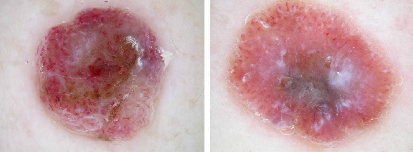
In the dermoscopic image of each lesion, we can see what is basically an atypical polymorphous vascular pattern and remnants of pigment.
We use the term atypical polymorphous vascular pattern to refer to the presence of 2 or more different vascular structures within a single lesion. Dotted vessels, linear-irregular vessels, and some hairpin vessels are visible in both our cases. The presence of an atypical polymorphous vascular pattern formed of dotted vessels and linear-irregular vessels has been found in 26.7% of amelanotic/hypomelanotic melanomas with a Breslow thickness of greater than 1mm.1 In addition, Menzies et al.2 discovered that this combination of vessels is predictive of melanoma, with an odds ratio (OR) of2.3.
Remnants of pigment are usually poorly defined areas of brownish, grayish-brown, or grayish-blue pigmentation that do not form clear dermoscopic structures (pigment network, globules, blue-gray veil, etc.). In the first case, the remnants of pigment have a very irregular distribution, whereas in the second case they are mainly found in the lower central area of the image. Pizzichetta et al.1 found irregular remnants of pigment in 86.7% of amelanotic/hypomelanotic melanomas with a Breslow thickness greater than 1mm, and Menzies et al.2 reported an OR of 2.6 for melanoma.
In view of the above, any neoplastic lesion that presents an atypical polymorphous vascular pattern and remnants of pigment must be excised and the diagnosis of amelanotic melanoma (or hypomelanotic melanoma, to be exact) should be included in the differential diagnosis. The lesions in both our patients were excised and the diagnosis was nodular amelanotic melanoma in both cases.
Analysis of the 2 images also reveals homogeneous whitish structures in an irregular distribution. Menzies et al.2 found these irregularly shaped areas of depigmentation to be one of the parameters most strongly predictive of melanoma (OR, 3.3).
Finally, there are dermoscopy structures called milky-red areas, which are usually poorly defined areas of pink (“milky-red”) discoloration that often contain vessels; these structures are highly suggestive of melanoma. Pizzichetta et al.1 found milky-red areas in 93% of amelanotic/hypomelanotic melanomas with a Breslow thickness greater than 1mm. The presence of these areas has an OR of 2.5 for melanoma according to Menzies et al.,2 and a positive predictive value of 78% according to Argenziano et al.3 Abundant milky-red areas are visible in both our cases, supporting the diagnosis of melanoma (Figs. 1 and 2).

Please cite this article as: Zaballos Diego P. ¡Ojo con los tumores rojos!. Actas Dermosifiliogr. 2014;105:872–873.


