


Animal-type melanoma is a rare variant of malignant melanoma in humans. Although many patients develop locoregional and distant metastases, mortality is lower than in other types of melanoma. We present 3 cases of animal-type melanoma in elderly men and include a description of common clinical and dermoscopic features (homogeneous blue pattern, irregular whitish structures, and irregular large vessels).
El melanoma de tipo animal es una variante infrecuente de melanoma maligno humano. A pesar de desarrollar metástasis locorregionales y a distancia, con frecuencia estos pacientes presentan una tasa de mortalidad inferior a la de otros tipos de melanoma. Presentamos 3 casos de melanoma de tipo animal en 3 varones de edad avanzada, con características clínicas y dermatoscópicas comunes (patrón azul homogéneo, estructuras blanquecinas irregulares y vasos gruesos irregulares).
Animal-type or equine melanoma is a rare variant of malignant melanoma. Histologically, it is characterized by heavily pigmented neoplastic cells with an epithelioid morphology and abundant cytoplasm throughout the dermis, with a scant or inexistent junctional component. Few cases have been published on animal-type melanoma and many aspects of its biologic behavior and prognosis are still unknown. Animal-type melanoma commonly metastasizes to locoregional lymph nodes and organs, but it appears to have a better prognosis than other types of melanoma.
We present 3 cases of animal-type melanoma diagnosed in our department. We describe the clinical, pathologic, and dermoscopic features observed and review the cases published to date.
Case DescriptionsCase 1A 74-year-old man with a history of prostate adenocarcinoma who presented with a 15-mm bluish-black nodule in the left parietal region that had been present for 2 years (Fig. 1A). Physical examination revealed multiple bluish papules measuring 2mm around the larger lesion. Dermoscopic evaluation showed a homogeneous blue pattern in all the lesions in addition to central ulceration and irregular whitish structures in the large nodule (Fig. 1B). The nodule and several of the satellite lesions were removed by excisional biopsy. The histopathology reports for both types of lesion showed a melanocytic proliferation throughout the dermis composed of heavily pigmented dendritic and epithelioid cells, accompanied by numerous melanophages (Fig. 1C). The findings were consistent with a diagnosis of animal-type melanoma. The maximum thickness (Breslow depth) was 4mm. Cerebral magnetic resonance imaging and a computed tomography (CT) scan of the thorax, abdomen, and pelvis showed no evidence of distant spread. The nodule and the surrounding lesions were excised with wide margins. Sentinel lymph node biopsy was not performed. Seventy months after diagnosis, the patient is free of disease and comes to the clinic for periodic follow-up.
.")
A Pigmented nodule in the left parietal region with multiple satellite lesions in the form of bluish papules. B, Dermoscopic image showing a homogeneous blue pattern, irregular whitish structures, and ulceration. C, Histologic image of the central part of the tumor with clusters of heavily pigmented epithelioid cells in the dermis (hematoxylin-eosin, original magnification ×40).
A 75-year-old man with a history of stroke secondary to severe atherosclerosis and carotid stenosis consulted for a 14-mm dark blue nodular lesion in the right parietal region (Fig. 2A). Dermoscopic features included a homogeneous blue pattern, ulceration in the lower part of the lesion, irregular whitish structures, and several large vessels (Fig. 2B). The histologic findings were compatible with a diagnosis of animal-type melanoma, with a Breslow depth of 3.2mm and a Clark level of iv. Melanoma was detected in the sentinel node in the right cervical lymph node region and we therefore performed a regional lymphadenectomy. No metastatic foci were found in any of the other lymph nodes. Sixty-six months after diagnosis, the patient is alive and there is no evidence of tumor spread.
Case 3
An 86-year-old man with a history of prostate adenocarcinoma presented with a 20-mm bluish-black nodule on his right arm (Fig. 3A). Dermoscopic features included a homogeneous blue pattern (the only feature of a melanocytic lesion), irregular whitish structures, and several large vessels (Fig. 3B). The histologic diagnosis was animal-type melanoma, with a Breslow depth of 5mm and a Clark level of v. A CT scan of the thorax, abdomen, and pelvis showed metastatic lesions (which were histologically confirmed) in the lungs and liver. The patient died of pneumonia 6 months later.
Discussion
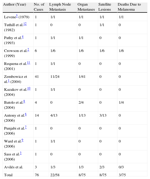
Animal-type melanoma has been referred to in the literature as heavily pigmented malignant melanoma, equine melanoma, and pigmented epithelioid melanocytoma. The term equine melanoma was coined because of the similarity between this variant of melanoma and the slowly progressive—and usually benign—tumor seen in gray horses. Zembowicz et al.1 used the term pigmented epithelioid melanocytoma to describe the tumor because of its similarity to epithelioid blue nevus. By using this term they were suggesting that animal-type melanoma was not a variant of malignant melanoma, but rather an independent entity encompassing both animal-type melanoma and epithelioid blue nevus. The literature contains descriptions of many lesions that could be considered animal-type melanoma. The most relevant cases are shown in Table 1.
Cases of Animal-Type Melanoma Reported in the Literature.
| Author (Year) | No. of Cases | Lymph Node Metastasis | Organ Metastases | Satellite Lesions | Deaths Due to Melanoma |
| Levene5 (1979) | 1 | 1/1 | 1/1 | 1/1 | 1/1 |
| Tuthill et al.12 (1982) | 1 | 0 | 0 | 1/1 | 0 |
| Pathy et al.4 (1993) | 1 | 1/1 | 1/1 | 0 | 0 |
| Crowson et al.2 (1999) | 6 | 1/6 | 1/6 | 1/6 | 1/6 |
| Requena et al.11 (2001) | 1 | 1/1 | 0 | 0 | 0 |
| Zembowicz et al.1 (2004) | 41 | 11/24 | 1/41 | 0 | 0 |
| Kazakov et al.10 (2004) | 1 | 1/1 | 0 | 0 | 0 |
| Batolo et al.8 (2004) | 4 | 0 | 2/4 | 0 | 1/4 |
| Antony et al.6 (2006) | 14 | 4/13 | 1/13 | 3/13 | 0 |
| Punjabi et al.7 (2006) | 1 | 0 | 0 | 0 | 0 |
| Ward et al.9 (2006) | 1 | 1/1 | 0 | 0 | 0 |
| Sass et al.3 (2006) | 1 | 0 | 0 | 0 | 0 |
| Avilés et al. | 3 | 1/3 | 1/3 | 2/3 | 0/3 |
| Total | 76 | 22/58 | 8/75 | 8/75 | 3/75 |
Animal-type melanoma has been described in patients of all ages, although the majority of cases have been in young adults.2 There does not appear to be a predilection for sex. Animal-type melanoma is more common in black and Hispanic individuals than other types of melanoma.1,3
Clinically, it usually presents as a well-circumscribed bluish-black nodule or plaque, although satellite lesions are relatively common.4,5 It is predominantly located on the scalp and the limbs. It generally appears de novo, and no association has been found with prior congenital or acquired melanocytic nevi, although there has been a report of a case occurring in association with a blue nevus.4 The association between animal-type melanoma and other predisposing factors, such as a family history of melanoma or sunburn, appears to be anecdotal.6
The biologic behavior of animal-type melanoma is unpredictable, and both regional lymph node and distant metastases are seen in many cases.2,6,9,10 Mortality rates, however, are lower than in other types of melanoma.2,5,10
Histologic features of animal-type melanoma include heavily pigmented dendritic and epithelioid cells that occupy the full thickness of the dermis and occasionally extend into the hypodermis.1,2,11 There are 2 types of cell populations: heavily, uniformly pigmented cells that tend to cluster in cords or sheets and less heavily pigmented cells with fine granular deposits in their cytoplasm. Melanophages may or may not be present. A junctional component may be seen and there has even been a report of intradermal animal-type melanoma.7 Reactive epidermal hyperplasia is relatively common. Other histologic features that may be observed are necrosis, ulceration, and features of regression.2,6,11
Nodular melanosis following complete regression of a melanoma can present challenges in the histologic differential diagnosis. To distinguish between neoplastic animal-type melanoma cells and melanophages, an immunohistochemical study (with S-100 protein, Melan A, or SOX-10) can be performed following whitening of the lesion with hydrogen peroxide or similar.
The histologic differential diagnosis should include other melanocytic lesions such as malignant blue nevus, deep penetrating nevus, melanoma metastasis, certain Spitz nevi, nodular melanoma, and primary dermal melanoma. Histologic diagnosis of animal-type melanoma is difficult, even for pathologists with extensive experience in pigmented tumors.
The dermoscopic features of animal-type melanoma have not been previously described. The 3 cases described in this article shared the following features: a homogeneous blue pattern (corresponding to the presence of heavily pigmented melanocytic cells in the dermis), irregular whitish structures (areas of epidermal hyperplasia), and a polymorphous vascular pattern generally composed of large irregular vessels, reflecting the prominent vascular component present in these tumors. These dermoscopic features, however, are not specific to animal-type melanoma and cannot therefore be used to differentiate between this type of melanoma and any other dermal melanocytic proliferations. Correlation of dermoscopic and histologic findings, however, may be useful in difficult-to-diagnose cases.
We have presented 3 cases of animal-type melanoma, a rare variant of melanoma, in 3 elderly men. Although all 3 patients developed metastasis, the outcome was favorable in 2 cases. We have also described dermoscopic features common to all 3 patients (a homogeneous blue pattern, irregular whitish structures, and thick vessels). Larger studies are necessary to confirm the specificity and diagnostic value of these features.
Ethical DisclosuresProtection of humans and animalsThe authors declare that no tests were carried out in humans or animals for the purpose of this study.
Confidentiality of dataThe authors declare that they have followed their hospital's protocol on the publication of data concerning patients and that all patients included in the study have received sufficient information and have given their written informed consent to participate in the study.
Right to privacy and informed consentThe authors obtained informed consent from the patients and/or subjects referred to in this article. This documentation is held by the corresponding author.
Conflicts of InterestThe authors declare that they have no conflicts of interest.
Please cite this article as: Avilés-Izquierdo JA, Leis-Dosil VM, Lázaro-Ochaita P. Melanoma de tipo animal: características clínicas y dermatoscópicas de 3 casos. Actas Dermosifiliogr. 2014;105:186–190.


