


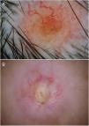
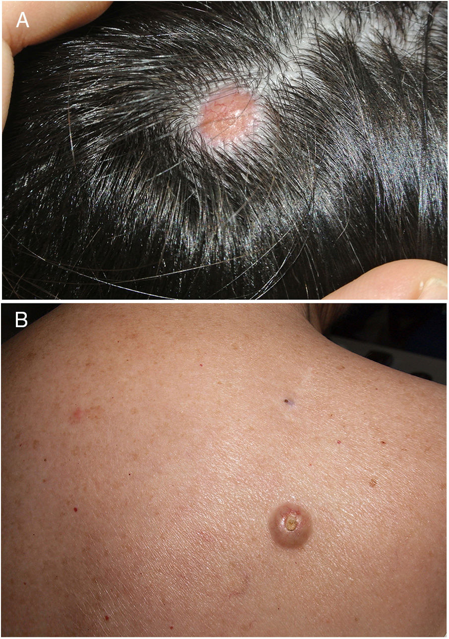
We present 2 cases that had previously been diagnosed as basal cell carcinoma based on the presence of arborizing vessels in dermoscopy. The first involved a 49-year-old woman, in whom the tumor was located on the scalp (Fig. 1A), and the second a 70-year-old woman, in whom the tumor was located in the area of the right scapula (Fig. 1B).
[[?]]What is your diagnosis?
CommentIn the first case, dermoscopy revealed arborizing vessels on a flesh-colored background with peripheral nonspecific brown areas (Fig. 2A). The patient was receiving treatment for disseminated carcinoma of the breast. Biopsy confirmed the definitive diagnosis of skin metastasis resulting from mammary carcinoma.
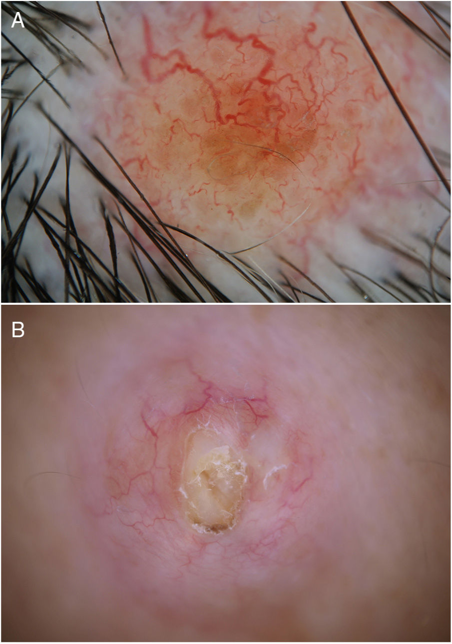
In the second case, dermoscopy revealed peripherally distributed arborizing vessels surrounding a central ulcerated area with a central yellowish background (Fig. 2B). Pressure released keratinizing material, thus confirming the diagnosis of infundibular epidermal cyst.
Arborizing vessels, also known as telangiectasias or “tree-like” vessels, are defined morphologically as large-caliber structures that are intensely red in color and branch into finer secondary vessels.
This type of vessel is one of the most characteristic dermatoscopic criteria of basal cell carcinoma.1
The literature contains numerous cases of lesions that mimic basal cell carcinoma owing to the presence of arborizing vessels. These include various types of adnexal tumors2 and infundibular cysts,3 as in the second case we report.
A study on the presence of arborizing lesions in conditions other than basal cell carcinoma revealed that up to 46% of lesions with arborizing vessels in dermoscopy were not basal cell carcinoma.4 These lesions included various cystic tumors (infundibular cyst, digital mucous cyst, milium), noncystic tumors (dermatofibroma, hypertrophic scars, intradermal nevi, and xanthogranulomas), and, albeit less frequently, nontumoral lesions such as necrobiosis lipoidica and morphea.4
The presence of arborizing vessels has also been described in some cases of skin metastasis from visceral tumors, such as those affecting the colon or ovary.5 Skin metastases from breast cancer often present pigmented structures that mimic melanoma more than basal cell carcinoma.6
While dermoscopy is very useful for increasing diagnostic accuracy, excessive attention to detail can sometimes present us from seeing the whole picture, as in the 2 cases we report. In no cases is the presence of a single dermatoscopic structure sufficient to confirm a diagnosis. Therefore, the presence of arborizing vessels in dermoscopy of a lesion should point us to other diagnoses in addition to basal cell carcinoma and to alternative clinical or dermatoscopic findings that help to establish a diagnosis.
Please cite this article as: Mateos-Mayo A, et al. Cuando los árboles no dejan ver el bosque. Actas Dermosifiliogr. 2020;111:159–160.


