


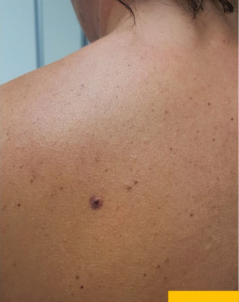
A 43-year-old woman with no relevant personal or family history of illness was referred by her primary care physician for evaluation of a pruriginous, violaceous lesion on the back of her trunk. The lesion had appeared 2 weeks earlier, and melanoma was suspected. She reported having noticed spontaneous bleeding from the lesion.
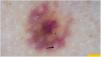
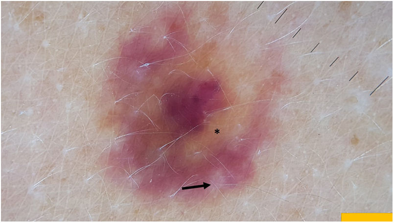
Physical examination revealed a violaceous papule surrounded by a purpuric halo (Fig. 1). Dermoscopy showed a structure with a reddish-purple center with lighter-appearing areas; the center was surrounded by a yellowish orange halo and another homogenous reddish halo at the periphery. Hair follicles were intact (Fig. 2).
Targetoid hemosiderotic hemangioma (THH).
CommentSanta Cruz and Aronberg first described a THH lesion in 1988, according to Sahim et al.1 THHs are described as benign vascular lesions that develop on the trunk and legs in middle age (around 30–40 years of age). Although the cause is still unknown, it has been suggested that a THH might be induced by trauma, specifically injury to a preexisting hemangioma.2
The presence of estrogen hormone receptors demonstrated by immunohistochemistry suggests that this hormone may accelerate endothelial proliferation and dilation, explaining the clinical behavior of THHs.3 D2-40 positivity and negative immunostaining for CD34 in endothelial cells, along with an absence of actin-positive pericytes, suggest that these lesions may have a lymphatic origin.4
A THH presents clinically as a reddish-violaceous papule surrounded by a violaceous, ecchymotic halo. The lesion may or may not be itchy or painful and is usually solitary. However, patients with multiple lesions have also been described.
THHs have a highly characteristic dermoscopic pattern, as shown in Fig. 2. In a study of 35 cases, the largest series published to date, the most common dermoscopic finding was of a dark reddish lacuna at the center and a reddish-violaceous homogeneous area around the periphery; that pattern was seen in 71.4% of the lesions.5 Our patient’s THH had a clearly biphasic halo around its center, a finding that defines the targetoid aspect of this lesion. The differential diagnosis includes consideration of malignant pigmented lesions such as melanoma,6 dermatofibromas, Kaposi sarcoma, insect bites, and basal cell carcinoma.7
THHs are benign vascular lesions that can be distinguished from others by their characteristic dermoscopic pattern. If the diagnosis is in doubt, in the absence of an ecchymotic halo, surgical removal and histopathologic study of the lesion would be the most prudent course of action.
Conflicts of InterestThe authors declare that they have no conflicts of interest.
Please cite this article as: Navarro-Triviño FJ. Pápula violácea en tronco. Actas Dermosifiliogr. 2021;112:647–648.


