


The journal Nature1 recently published an article featuring an image (Fig. 1) that immediately caught our attention. If the image was shown in a Rorschach test, a virologist would probably say “coronavirus”, but a dermatologist trained in dermoscopy might well say “a growing melanocytic nevus” or even “melanoma”.
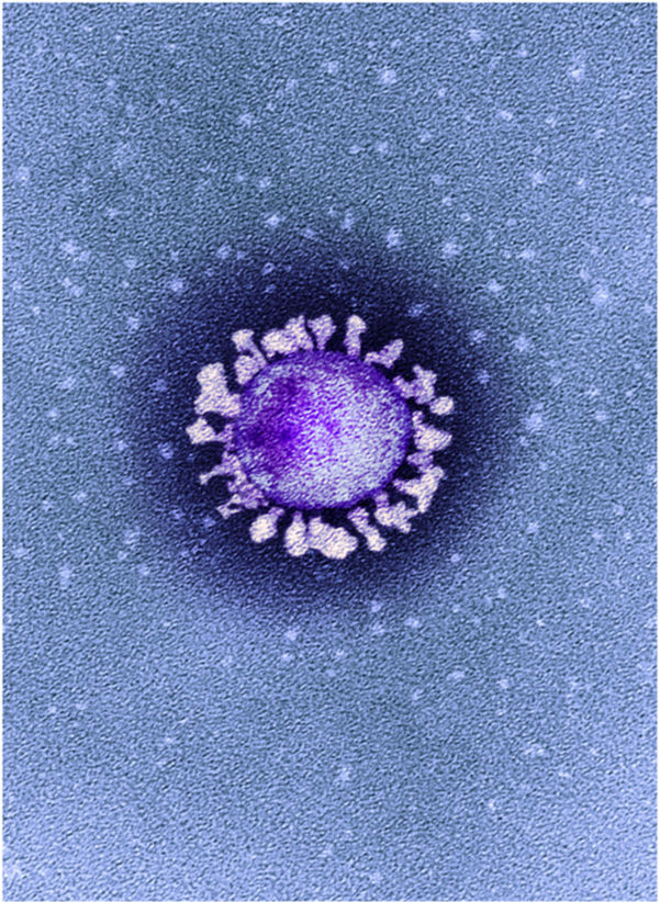
Colored transmission electron micrograph showing a SARS-CoV-2 particle isolated from a patient with COVID-19 in the United Kingdom. ©National Infection Service, United Kingdom/Science Photo Library/agefotostock.
Note the characteristic outward projecting spikes or protuberances on the surface and the markedly irregular rim and somewhat heterogeneous morphology.
Several aspects of the image reminded us of dermoscopic features we have seen in dysplastic melanocytic nevi and incipient melanomas (Fig. 2).
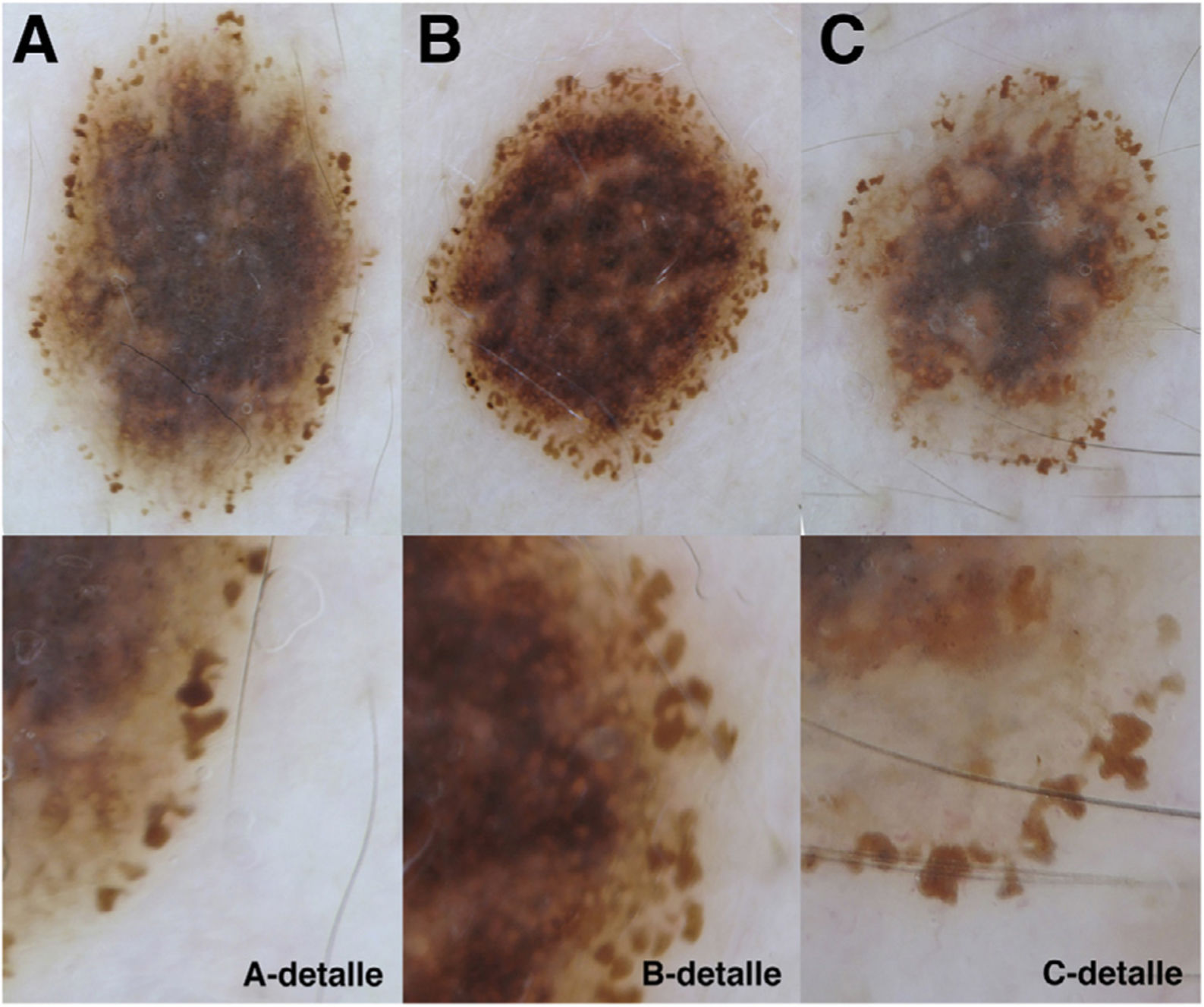
Polarized light dermoscopic images.
Lesions with large pigmented peripheral structures and an irregular morphology, similar to SARS-CoV-2 protein spikes. Detailed image of different areas of the lesions are shown in the panels underneath. A, C, Lower right corner. B, Right edge.
The histopathologic diagnoses were dysplastic nevus (A), melanoma in situ (B), and superficial spreading melanoma (C).
Dermoscopy reveals markedly irregular, large polymorphous peripheral structures, often long in shape and with an angulated rim, similar to the spike proteins of the virus particle in the electron micrograph in Fig. 1. These features are indicative of a growing atypical melanocytic lesion (dysplastic nevus or incipient melanoma).
CommentZalaudek et al.2 described the presence and significance of peripheral globules in melanocytic lesions more than a decade ago. These globules, which frequently project outwards from the silhouette of the lesion, and sometimes appear to be disconnected, usually indicate active growth.3,4 Many expanding junctional or compound nevi have a peripheral rim of pigmented globules, which are also common in the most active growth phase of Spitz or Reed nevi.5
Peripheral globules are occasionally seen in melanoma. In a recent dermoscopic study of the morphology and distribution of peripheral globules in melanocytic lesions, Reiter et al.6 suggested that lesions with asymmetrically distributed globules or globules with varying shapes, sizes, and colors should be biopsied or closely monitored–especially pigmented lesions on the extremities—as they may be signs of an incipient melanoma.
Reiter et al.6 did not include lesions with pseudopods in their study. While pseudopods are also round or oval structures that project outwards from the edge of lesions, they are not globules in the strict sense of the word (the last edition of the Merriam-Webster dictionary defines globule as "a tiny globe or ball especially of a liquid"). Similarly, we do not believe that the structures shown in Fig. 2—long, polymorphous, pigmented structures, some with an angulated rim—can be described as peripheral globules. Strictly speaking thus, they should not be called globules, nor attributed the same biologic or diagnostic significance as classic peripheral pigmented globules in melanocytic lesions.2–5
In our experience, these highly irregular pigmented peripheral structures are rare and are neither globules nor pseudopods. We have recently seen several cases of severely dysplastic melanocytic nevi and incipient melanomas in which the most distinctive and suspicious finding was a peripheral ring (sometimes incomplete) featuring structures of this type (Fig. 2).
In light of the electron micrograph shown in Fig. 1 and the fact that we have described these dermoscopic features during the COVID-19 pandemic, we have decided to call them SARS-CoV-2 spike-like pigmented peripheral structures. We believe that the morphologic similarities between the viral spike proteins and these structures are clearly depicted in Figs. 1 and 2. We do not believe that their biologic significance or diagnostic value should be likened to those of classic pigmented peripheral globules, although further studies are warranted to confirm their biological and prognostic significance.
Conflicts of interestThe authors declare that they have no conflicts of interest in relation to the content of this article.
Please cite this article as: Martin-Gorgojo A, Ramírez-Bellver JL, Ruiz-Rodríguez R, Pizarro Á. Estructuras pigmentadas periféricas de morfología similar a las espículas del SARS-CoV-2 como hallazgo dermatoscópico sugerente de nevo displasico y melanoma incipiente. Actas Dermosifiliogr. 2022;113:72–73.


