Información de la revista
Vol. 114. Núm. 5.
Páginas T435-T436 (Mayo 2023)
Compartir
Compartir
Descargar PDF
Más opciones de artículo
Visitas
1750
Vol. 114. Núm. 5.
Páginas T435-T436 (Mayo 2023)
Practical Dermoscopy
Acceso a texto completo
[Translated article] Progressively Appearing Papules on the Penis
Pápulas peneanas de aparición progresiva
Visitas
1750
A. Martin-Gorgojo
Autor para correspondencia
alejandromartingorgojo@aedv.es

Corresponding author.
, A. Comunión-Artieda, F.J. Bru-Gorraiz
Sección de Especialidades Médicas, Servicio de ITS/Dermatología, Ayuntamiento de Madrid, Madrid, Spain
Contenido relaccionado
Actas Dermosifiliogr. 2023;114:435-610.1016/j.ad.2022.05.027
A. Martin-Gorgojo, A. Comunión-Artieda, F.J. Bru-Gorraiz
Este artículo ha recibido
Información del artículo
Texto completo
Bibliografía
Descargar PDF
Estadísticas
Figuras (2)
Texto completo
Case Presentation

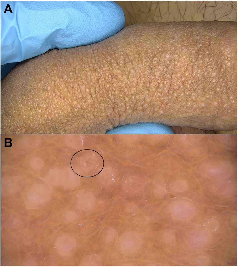
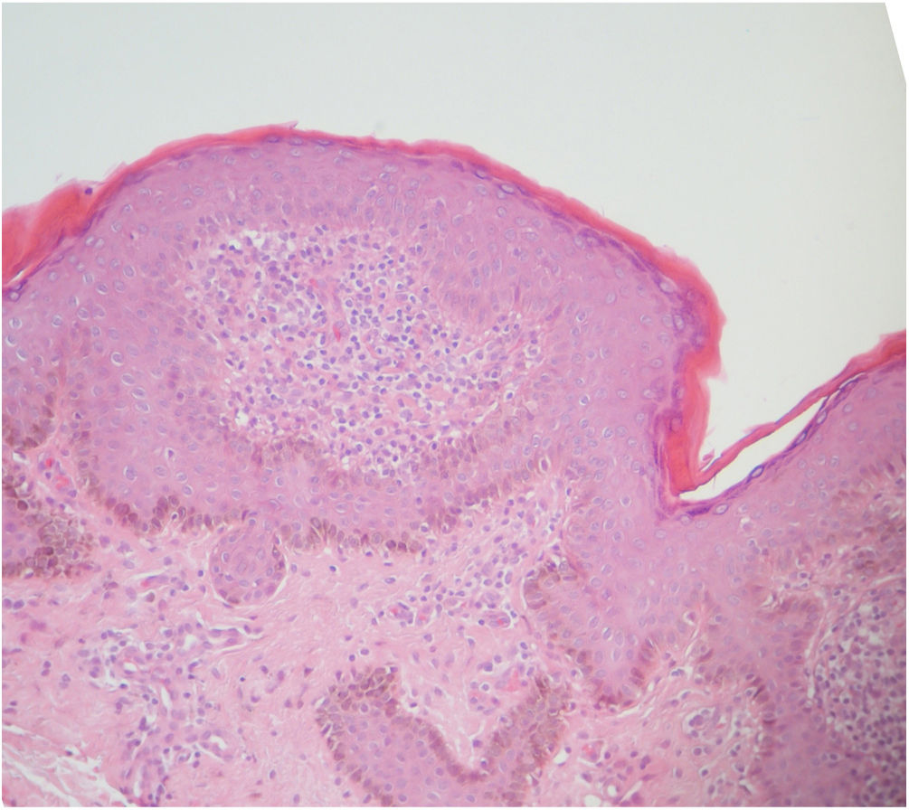
A 25-year-old man presented for evaluation of slightly itchy penile papules that had appeared progressively over the preceding year. Examination revealed numerous rounded, shiny, pale white papules on the shaft of the penis (Fig. 1A) and some isolated papules on the glans penis. No other similar lesions were observed. A skin biopsy was taken to confirm the diagnosis (Fig. 2).

Figure 1.

Case presentation. A, Clinical presentation. B, Dermoscopic findings (obtained using Fotofinder digital dermoscopy, 20× magnification).

(0,21MB).
Figure 2.

Punch biopsy histology (hematoxylin–eosin).

(0,14MB).
What is Your Diagnosis?

Lichen nitidus.

Comment

Lichen nitidus1 is an inflammatory process that manifests clinically with uniform, whitish or skin-colored, generally asymptomatic papules of 1–2mm in size. Dermoscopic examination reveals structureless white areas (corresponding to papules), often with a darker central spot, which tends to diminish or interrupt the overlying skin markings, and a characteristic “sunburst” pattern (skin markings that radiate from the papules) that is particularly visible with nonpolarized dermoscopy.2,3

The most important conditions to include in the differential diagnoses of lichen nitidus of the penis are Fordyce glands and Molluscum contagiosum lesions. Dermoscopy is a useful tool to distinguish between these 2 entities: Fordyce glands appear as yellowish lobules surrounded by vascular garland-like structures, while Molluscum contagiosum lesions appear as polyglobular, yellowish-white, amorphous structures with a peripheral crown of fine vessels.

Conflicts of Interest

The authors declare that they have no conflicts of interest.

References
[1]
X. Vilanova, C. Cardenal, J.M. Capdevila.
Liquen nitidus.
Actas Dermosifiliogr, 48 (1957), pp. 221-229
[2]
D. Jakhar, C. Grover, I. Kaur, S. Sharma.
Dermatoscopic features of lichen nitidus.
Pediatr Dermatol, 35 (2018), pp. 866-867
[3]
S. Neema, S. Goyal.
Dermoscopy of anserine folliculosis.
Dermatol Pract Concept, 11 (2021), pp. e2021019
Copyright © 2022. AEDV
Idiomas
Actas Dermo-Sifiliográficas
Opciones de artículo
Herramientas
es en

¿Es usted profesional sanitario apto para prescribir o dispensar medicamentos?

Are you a health professional able to prescribe or dispense drugs?