


Interpreting patch test reactions is not easy. It requires experience and is characterized by high intraobserver and interobserver variability. It can sometimes be truly difficult to discern between a weak allergic reaction and an irritant reaction. A number of recent studies have investigated the dermoscopic features of patch test reactions. Homogeneous erythema is the main feature observed in patients with a positive allergic reaction, although dotted vessels, vesicles, crusts and yellow-orange areas may also provide clues. These features are somewhat similar to those observed in inflammatory conditions, such as eczema. In patients with an irritant reaction, the most common dermoscopic findings are the pore reaction pattern and perifollicular erythema. Dermoscopy could be useful for establishing a diagnosis in the case of doubtful patch test reactions.
La lectura de los parches de pruebas epicutáneas no es sencilla, presenta una gran variabilidad inter- e intraobservador y requiere experiencia. En ocasiones es realmente difícil determinar si se trata de una reacción alérgica de intensidad leve o si estamos ante una reacción irritativa. Recientemente se han publicado algunos trabajos que han estudiado las características dermatoscópicas de las distintas reacciones que se producen tras realizar pruebas epicutáneas. La característica dermatoscópica más frecuentemente observada en los parches alérgicos es el eritema homogéneo, si bien vasos puntiformes, vesículas o costras y áreas amarillo-anaranjadas también parecen indicar una reacción alérgica, guardando cierta similitud con lo observado en patología inflamatoria como en el eccema. Por otro lado, en cuanto a las reacciones irritativas, el patrón más indicativo sería el «patrón del poro», acompañado de eritema perifolicular. Estos hallazgos dermatoscópicos pueden ser de utilidad al clínico en sutoma de decisiones ante una reacción dudosa.
In our setting, patch test reactions should be interpreted according to guidelines such as those of the European Society of Contact Dermatitis (ESCD),1 which classifies them as positive, negative, or irritant. Sometimes, the results are doubtful or weak positive and it is difficult to make a clear diagnosis, leading to marked interobserver variability. In these cases, the test is usually repeated to try to reach a reliable diagnosis, thus necessitating consumption of resources.
With the aim of setting more objective parameters that could help diagnosing doubtful cases, some authors suggested measuring erythema using software applications and scales.2,3 Nonetheless, these approaches have not gained widespread approval owing to technical requirements, the lack of universal availability, and the fact that they are time-consuming and do not provide a clear-cut answers.
Dermoscopy is a simple, rapid, harmless, and noninvasive technique that has been widely used for the diagnosis and follow-up of patients with skin cancer and various inflammatory diseases.4 However, application of this approach for evaluation of patch test reactions in the diagnosis of allergic contact dermatitis (ACD) is very limited. Early studies reported the dermoscopic characteristics of allergic and irritant patch test reactions in an attempt to establish differential criteria.5–7 However, since the authors restricted their choice to patch tests with an unequivocal clinical diagnosis, only general recommendations could be made. In 2021, Oppermann et al.8 published the results of the first study to specifically use dermoscopy for doubtful and weak positive reactions and provided dermoscopic keys and patterns that could improve diagnostic capacity in these cases.
In the present study, we review all the available literature to date on the use of dermoscopy for the interpretation of patch test results specially pointing out the dermoscopic criteria that could help diagnosing doubtful cases.
Dermoscopy in Allergic ReactionsIn clinical terms, patch test reactions indicating contact allergy are classified into 3 levels according to the ESCD as follows: weak positive (+), manifesting as erythema and infiltration that may or may not be associated with papules; strong positive (++), if papules and vesicles are also present; and extreme positive (+++), with intense erythema, infiltration, and coalescing vesicles.1
Corazza et al.5,6 described the dermoscopic characteristic of allergic patch tests for the first time in 2 studies in which the patch test reactions were unequivocally clinically diagnosed as allergic or irritant. Erythema was the most characteristic finding and was almost always present in both extreme positive reactions (++/+++) and in weak positive reactions (+). This finding was significantly more frequent in allergic reactions than in irritant reactions.5–7 Although Opperman et al.8 did not evaluate the intensity of the erythema, although they did assess its distribution and identified a homogeneous and diffuse erythema pattern occupying an area>50% in more than 90% of allergic reactions (Fig. 1a2). Therefore, they suggested that the presence of homogeneous and diffuse erythema was necessary to establish a diagnosis, irrespective of the intensity of the erythema and involvement of the follicular areas. In our experience, homogeneous and diffuse erythema that is not limited to the follicular areas and covering more than half of the chamber could be the main dermoscopic finding when differentiating between an allergic test result and an irritant one.

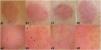
Allergic reactions (++/+++). Upper row (1), clinical images; lower row (2), dermoscopic images. All the dermoscopic images reveal homogeneous and diffuse erythema, especially in a2, but also in (b2), “soap bubble” vesicles (black arrowheads), (c2) dotted vessels, occasional crusts, and yellow-orange areas, and (d2) nonfollicular pustules (green arrowheads).
Initial studies reported whitish “soap bubble” vesicles (Fig. 1b2) as findings that were highly prevalent, sensitive, and specific for allergic reactions5,6 and that were found not only in more or less extreme allergic reactions (++/+++),7,8 but were also reported to be the main characteristic that made it possible to differentiate between weak reactions (+) and irritant reactions.5,6 The finding was attributed to the potential for spongiosis to lead to the formation of vesicles and exudate that would not be clinically visible but would be visible using dermoscopy. However, and in line with our experience, other authors did not find these structures in weak positive reactions.8
Corazza et al.5,6 found vascular structures to be highly sensitive and prevalent in allergic reactions and to be constant in weak positive reactions, with a negative predictive value of 100%, suggesting that the absence of such reactions would almost rule out a diagnosis of allergic reaction.6 In contrast, other studies only report vascular abnormalities that can be distinguished from basal erythema in little more than 20% of cases (our experience is similar).8 As for morphology, dotted vessels (Fig. 1c2) were strongly associated with allergic reactions, in terms of both prevalence and number.6 The authors suggest that this association is probably due to the inflammatory nature of the reaction, similar to findings for dermatoses such as eczema and psoriasis. It is noteworthy that other vascular structures have been identified in allergic reactions, including glomerular and petechial vessels, which, as with dotted or linear vessels, are not specific and whose presence does not rule out the diagnosis. In our experience, few patch tests were characterized by vascular abnormalities in dermoscopy, and the number of cases found in doubtful or weakly positive patches is limited. Based on these data, we suggest that the presence of dotted vessels, which matches dermoscopy findings for eczema, would support a diagnosis of ACD, although the absence of such findings or the presence of other types of vessel would not rule out this diagnosis.
Lastly, the orange-yellowish areas or crusts (Fig. 1c2) observed in allergic reactions correspond to the dermoscopic image of dry exudate in acute eczema.4,8,9 This finding is very suggestive of allergic reaction,5,6,8 even in weak reactions, although it is only present in 21.3%–35% of cases, where they are less evident and less frequent in our clinical practice.
Furthermore, we may also observe other, less specific characteristics of allergic reactions, such as papules, pustules (Fig. 1d2), perifollicular erythema, and pore reaction. These characteristics could be more indicative of an irritant reaction, especially pore reaction, although they do not rule out the possibility of an allergic reaction. Opperman et al.8 pointed out that, if any of these characteristics, they should be accompanied by a diffuse and homogeneous erythema before a diagnosis of allergic reaction can be confirmed.
Dermoscopy in Irritant ReactionsIn clinical terms, irritant reactions are characterized by the “burnt” appearance of the skin, with cigarette paper texture, typically without erythema (or scant erythema) and greater or lesser associated follicular reaction.1
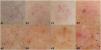
The dermoscopic characteristics of irritant reactions are less defined than those of allergic reactions, probably because they are associated with a wide range of epidermal changes.5,6 In an attempt to define them, Corazza et al.6 performed patch tests with sodium lauryl sulfate 2.5% applied over 48hours as a positive control for irritant tests but found no clinical differences with irritant reactions in daily clinical practice. The authors described erythema as being less intense than in allergic reactions. However, in a subsequent study, they found no differences between the mean score for erythema in extreme irritant reactions in controls and weak allergic reactions; therefore, the intensity of erythema cannot be used to differentiate between them.6 With respect to this characteristic, other authors report that the distribution of the erythema is more important than its intensity, with nonallergic reactions defined as those without diffuse erythema, even if they involve isolated perifollicular erythema and follicular crusts are present.8 Therefore, they describe the dermoscopic characteristics of irritant reactions as pore reaction and/or perifollicular reaction, in the absence of homogeneous basal erythema. The”poral reaction” (Fig. 2a2), described by Yang et al.10 as a diffuse dotted brown pigment deposit varying in size and surrounded by a yellowish halo corresponds to the dermoscopic equivalent of the”poral” reaction (Fig. 2a1) described by Storrs and White11 and which is the result of a toxic effect of cobalt on the acrosyringium.
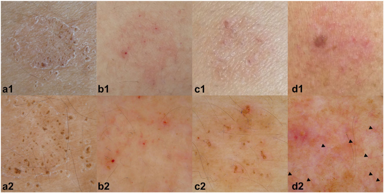
As for vascularization, while it was initially suggested that this would be less evident in irritant reactions than in allergic reactions,5 subsequent studies reported that the mean scores for the vessels did not differ.6 However, since the mean value of the dotted vessels was significantly lower for irritant reactions, their absence could point more towards this type of reaction. In any case, no clear pattern of specific vascular changes has been defined in irritant reactions, although some studies report a greater percentage of linear vessels (Fig. 2d2) than dotted vessels, with no significant differences between them.8 Consistent with these findings, our experience suggests that linear vessels would be more suggestive of irritant reactions.
Lastly, although vesicles have been reported to be characteristic of allergic reactions,6 they are also found in up to 9% of irritant reactions, according to some studies, possibly owing to a spongiosis phenomenon or irritant cytolysis that would be visible both in histology12 and in optical coherence tomography.13
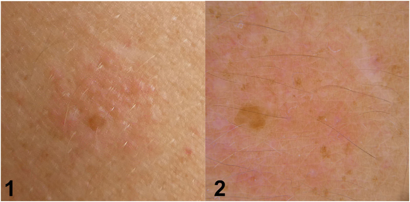
Dermoscopy in Clinically Doubtful ReactionsAccording to the ESCD, doubtful reactions are those that present with faint erythema only, which cannot be classified as irritant, since it is not predominantly perifollicular. Furthermore, as these reactions are not infiltrated, they are not palpable.1 However, in daily clinical practice, they are often difficult to identify, since we may find patches that, while palpable, are not characterized as having clinical erythema. Dermoscopy could aid decision making in such cases.
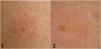
Only 1 article has focused on the characteristics of doubtful reactions. The authors stress that dermoscopy helps to better define erythema, enabling up to 91.6% of doubtful or weak positive reactions to be reclassified.8 Reactions reclassified as positive were almost always characterized by diffuse and homogeneous erythema occupying more than 50% of the chamber. Patch findings diagnosed as negative in dermoscopy do not present such homogeneous erythema, even with perifollicular erythema and/or crusts. Lesions in which the presence of a nonfollicular erythematous area could not be determined were diagnosed as dermoscopically doubtful (Fig. 3).
Unlike Corazza et al.,5,6 Opperman et al.8 did not find vesicles in doubtful or weak positive reactions or dotted vessels. They only found linear vessels in 3 of 44 cases and polymorphic vessels in 1 of 44 cases. In our clinical practice, we did not usually find vessels in addition to erythema in doubtful or weak positive reactions, or we were unable to classify the findings. However, the presence of dotted vessels pointed us more toward an allergic reaction.
In our experience, dermoscopy has proven useful for evaluating the presence and pattern of erythema in doubtful lesions, in which it is not clearly clinically evident. This is probably the scenario where this approach becomes most useful. As with Opperman et al.,8 diffuse and homogeneous erythema occupying more than half of the patch is an almost indispensable characteristic that could be accompanied by other more or less specific findings of an allergic or irritant reaction, without these playing a major role in diagnosis. Table 1 summarizes and compares dermoscopic findings in allergic and irritant reactions.
Dermoscopy Findings in Allergic and Irritant Reactions.
| Dermoscopic characteristic | Allergic reaction | Irritant reaction |
|---|---|---|
| Erythema | Diffuse and homogeneous pattern>50% | Perifollicular pattern |
| Vesicles | ↑↑↑ | ↑ |
| Pustules | ↑ | ↑↑ (follicular) |
| Yellow-orange crusts | ↑↑↑ | ↑ |
| Pore pattern | ↑ | ↑↑↑ |
| Dotted vessels | ↑↑↑ | ↑ |
| Linear vessels | ↑ | ↑↑↑ |
↑↑↑ Very frequent finding, ↑↑ moderately frequent finding, ↑ infrequent finding.
In the case of patch tests with dyes or in patients with dark skin phototypes, dermoscopy is a key element when evaluating homogeneous erythema, since it might not be clinically evident, and some substances might leave pigment deposition (Fig. 4).7,8 In our experience, dermoscopy has made it possible to observe true erythema in a patch test with dyes which would be clinically difficult in clinical terms, since it is possible to distinguish the particles of the substances tested on this erythema, which, in many cases mimic it or prevent the correct visualization of the erythema by settling on top of it.
Conclusion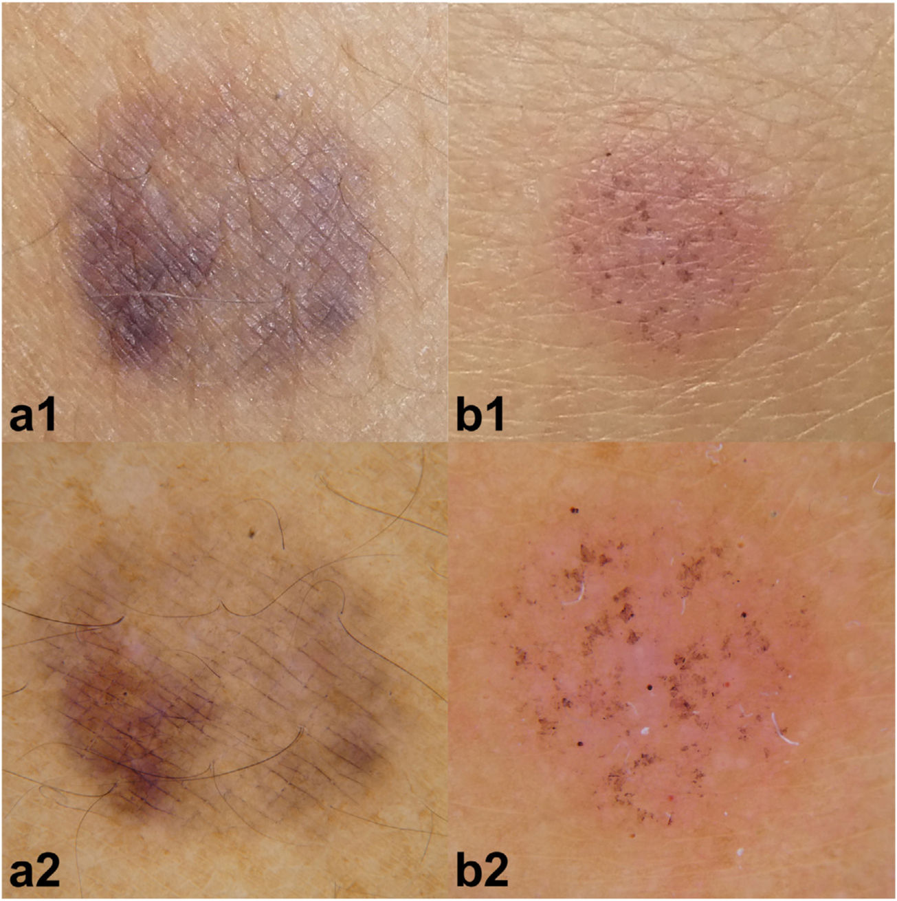
Dermoscopy has become an additional tool for the evaluation of patch tests and has proven very useful in the interpretation of weak reactions and, moreover, in doubtful reactions. Thus, it also enables better definition of dermoscopic characteristics, especially erythema, in reactions in patients with dark skin phototypes and in reactions caused by substances with pigment deposition.
Despite differences between the various studies, in general, allergic reactions are characterized by the almost constant finding of diffuse and homogeneous erythema—to the extent that its absence would lead us to question the diagnosis—and by findings such as yellow-orange crusts and vesicles, in line with several findings expected in acute eczema. Irritant reactions are often characterized by the “poral pattern” or perifollicular erythema. Therefore, all those lesions where erythema is not limited to the follicle and that cannot be classed as clearly diffuse and homogeneous in >50% of the chamber, with no other structures present, would be classified as doubtful reactions. Other reported structures, such as, dotted vessels and linear vessels, have not been significantly associated with allergic reactions or irritant reactions; therefore, they would not affect the diagnosis. Nevertheless, since their presence has been reported to be more characteristic of either one of the reactions, further studies are needed to determine their true diagnostic value.
Conflict of InterestThe authors declare that they have no conflicts of interest.


