


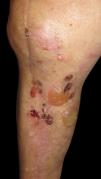
A 79-year-old male was referred from the traumatology department due to the appearance of pruritic blisters on the right knee, 8 weeks after undergoing total knee arthroplasty.
Physical examinationPhysical examination revealed the presence of tense serohemorrhagic blisters and crusts located around the surgical scar, without lesions elsewhere (Fig. 1).
Supplementary tests
Bacterial culture turned out negative. Patch testing performed using the Spanish standard series (GEIDAC), metals, methacrylates, benzoyl peroxide, vancomycin, and gentamicin, all turned out negative as well.
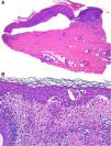
HistopathologyTwo skin biopsies were performed. The first showed a subepidermal blister with scant inflammatory content (Fig. 2A), and the second marked eosinophilic spongiosis (Fig. 2B).

What is your diagnosis?
DiagnosisThe diagnosis was localized bullous pemphigoid.
Differential diagnosisDifferential diagnosis in the presence of postoperative blistering lesions includes bullous impetigo, which was ruled out by the negative culture, and allergic contact eczema, excluded based on patch testing. In case of a histological finding of a subepidermal blister with minimal inflammatory infiltrate, other considerations include the cell-poor variant of bullous pemphigoid, epidermolysis bullosa, porphyria cutanea tarda, burns (e.g., from cryotherapy), and even toxic epidermal necrolysis.1 The clinical picture, along with the second biopsy showing intense eosinophilic spongiosis, led us to the diagnosis of localized bullous pemphigoid,2 which was confirmed after detection of anti-basement membrane antibodies in the patient's serum.
CommentaryLocalized bullous pemphigoid is considered a rare variant of the disease,3,4 often overlooked and likely underdiagnosed in clinical practice.4 However, it is important to consider it in the differential diagnosis, especially when preceded by triggering factors. Diagnosis is based on clinical presentation and additional tests, such as biopsy (with compatible DIF and/or positive IIF in serum), sharing criteria with the classic form of the disease, as it lacks specific diagnostic criteria.3 Various triggers have been described, including burns, radiotherapy, and a variety of surgical procedures—among which orthopedic procedires, particularly arthroplasties, are notable.2,5,6 In most cases, DIF is positive, although in was negative our patient. Conversely, IIF in serum is usually negative. The presence of serum antibodies is considered a specific marker of the disease and has been correlated with its severity; hence, in localized forms, it may remain negative due to the self-limiting nature of the disease.3,4 The latency period from surgery to the onset of lesions varies among studies, although it typically appears weeks after surgery, as in our case.5 Although the causal mechanism is unknown, it is hypothesized to be due to an isotopic phenomenon, caused by disruption of the dermoepidermal junction during surgery, leading to a local immune imbalance with autoantibody formation that ultimately results in subepidermal blistering.2,4
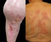
Disease progression and treatmentAfter a brief regimen of oral and topical corticosteroids, the lesions and itching resolved completely; however, upon tapering the dose, the patient experienced another outbreak (Fig. 3A). Six weeks after the first flare, new urticarial-like lesions appeared on the neck, trunk, and arms (Fig. 3B), without epidermal detachment and accompanied by intense pruritus. Histologically, these lesions showed eosinophilic spongiosis similar to the knee lesions, consistent with secondary generalization of bullous pemphigoid. Although the generalization of localized bullous pemphigoid has been reported, it is a rare finding.4 The patient required a prolonged 8-month regimen with oral doxycycline, along with intermittent short regimens of oral corticosteroids during relapses, with no new recurrences to date at the >1-year follow-up.
Authorship
The authors contributed directly to the intellectual content of this manuscript (Dr. Giulia Greta Dradi, Dr. Enrique Gómez de la Fuente, and Dr. Elena García Zamora), the genesis and analysis of data (Dr. Giulia Greta Dradi), and approve its contents submitted for editorial review, giving consent for their names to appear as authors.
FundingNone declared.
Conflict of interestThe authors declare that they have no conflict of interest.


