


Anterior hairline measurements and their possible relationship with androgen levels, sebum production, and skin hydration have not been reported in white Spanish women.
Material and methodsThis was a prospective descriptive and analytical observational study conducted on 103 healthy premenopausal white Spanish women recruited from the health staff of Hospital Universitario Virgen Macarena in Seville, Spain. Measurements were made of anterior hairline implantation, sebum levels, and the degree of hydration of the stratum corneum. Androgen levels were also determined in 50 volunteers from this group 3 to 5 days after the end of the menstrual cycle.
ResultsThe mean age of the women was 29.7 years. A widow's peak was observed in 94.17% of the group. The mean dimensions of the widow's peak were a height of 1.01 cm and width of 2.13 cm. The mean hormone levels were within normal limits for our hospital's laboratory with the exception of 17-hydroxyprogesterone, with a mean level of 1.39 ng/mL (range, 0.6-5.9ng/mL; normal limits, 0.15-1.10 ng/mL). The mean prostate specific antigen level was 0.04ng/mL (range 0.02-0.08ng/mL; normal limits, 0.00-0.02ng/mL).
ConclusionsThe hairline measurements of the white Spanish women in this study differ from those reported in American women. Knowledge of this normal pattern of anterior hairline implantation can be important in the evaluation of women with female androgenetic alopecia with male pattern, frontal fibrosing alopecia, or other established scarring alopecia seeking a surgical solution by hair transplantation.
Las medidas de la línea de implantación pilosa frontal de la mujer española caucásica no han sido descritas, y tampoco si existe relación entre estas medidas y los niveles hormonales androgénicos, producción sebácea e hidratación.
Material y métodosEstudio observacional, prospectivo, descriptivo y analítico en 103 mujeres sanas españolas caucásicas premenopáusicas, pertenecientes a la plantilla sanitaria de nuestro hospital a las que se les midieron la línea de implantación pilosa frontal, los niveles de sebo y la hidratación de la capa córnea, y en 50 voluntarias de este grupo se les determinaron, además, los niveles hormonales androgénicos 3-5 días después de terminar la menstruación.
ResultadosLa edad media de nuestras pacientes fue de 29,7 años. El pico de viuda se objetivó en 94.17% de las pacientes. Las dimensiones medias del pico de viuda en nuestro grupo fueron de 1,01cm de alto y 2,13cm de ancho. Los niveles medios hormonales fueron los que habitualmente consideramos como normales en el Laboratorio de nuestro hospital excepto la 17-hidroxi-progesterona que alcanzó valores medios de 1,39 ng/mL (rango: 0.6-5,9; N: 0.15-1,10). El PSA fue de 0.04 ng/mL (rango: 0.02-0.08; N: 0.00-0.02).
ConclusionesLas medidas de las líneas de implantación pilosa frontal de la mujer caucásica española han sido distintas de las descritas para la mujer americana. El conocimiento de este patrón de distribución de la línea de implantación pilosa frontal normal puede ayudar en el caso de que la paciente con una alopecia androgenética femenina de patrón masculino, una alopecia frontal fibrosante u otra cicatricial ya estable, desee corregir quirúrgicamente su problema mediante trasplante capilar.
Female alopecia may be either androgenetic or nonandrogenetic. In women, the androgenetic form manifests with miniaturization of the hair shafts and gives rise to 2 different patterns of alopecia.1 The first of these is female androgenetic alopecia, a pattern classified by both Ludwig2 and Olsen3 and characterized by a decrease in the diameter of the hair shaft—a process also known as rarefaction—and by diffuse hair loss that results in a triangular, alopecic area at the front of the vertex, but not by changes in the anterior hairline. The second pattern is androgenetic alopecia in women mimicking male pattern baldness, which was classified into 5 types by Ebling.4 This variant, characterized by thinning hair in the parietal and frontovertical scalp, is found in women with persistent adrenarche syndrome and ovarian or adrenal tumors. Occasionally, it develops after hysterectomy or forms part of age-related hair loss and it is differentiated from the female pattern in that the anterior hairline is affected.
The male pattern variant results in a receding hairline in women just as it does in men. Therefore, we need data on the normal contour and structure of the hairline so that an aesthetically appropriate result can be achieved when surgical reconstruction is required. This information is also needed for the treatment of the growing number of patients presenting with frontal fibrosing alopecia and other stable scarring alopecias that affect the anterior hairline.
Anterior hairline patterns and the different racial variants of alopecia in men were described many years ago5–9; however, the characteristics of natural female hairlines have not yet been determined. In 2009, Nusbaum and Fuentefria10 published the anterior hairline characteristics of 360 American women who were clients of hair salons. In 2011, Jung et al.11 identified 5 basic hairline shapes or patterns in 130 randomly selected Asian women.
A limitation these studies share is that the authors failed to set selection criteria obliging them to ascertain whether the participants (both those assessed in hair salons10 and those randomly selected11) had any dermatological or hormonal disorder that might influence the hairline dimensions measured. Moreover, given the existence of anthropometric differences related to race, the results obtained in American or Asian women may not be applicable to a Spanish population.
The aim of our study was to characterize the anterior hairline patterns of a sample of healthy white Spanish women aged between 18 and 45 years—focusing specifically on the frequency of occurrence of the widow's peak and the dimensions of the other structures that make up the hairline—and to determine the normal hormonal profile in this group. Furthermore, as increased sebum discharge is common in alopecia, we also measured the amount of oil secreted by normal white Spanish women and assessed skin hydration on the face and body.
Material and MethodsThis was a prospective, descriptive, observational analysis of a group recruited from the clinical staff of our hospital. The study was carried out between January and June 2010.
In total, 105 white Spanish women of childbearing age (18-45 years) were recruited prospectively from among the staff of our hospital. None of the participants had a concomitant systemic disease or clinical signs suggestive of underlying hormonal disorders (alopecia, acne, hirsutism, or menstrual disorders). We subsequently excluded 2 women who were 2 and 3 months pregnant, respectively, but who were unaware of their condition when they entered the study.
In an oral interview we collected the following data: age, age at menarche, skin phototype, hair and eye color, known history of iron deficiency, oral contraceptive use, and smoking status.
The following tests were performed on all the participants:
- 1.
Sebumetry: we measured skin surface sebum on the face (forehead) and body (flexor surface of the left forearm) with a sebumeter (Skin Diagnosis SD 27, Courage + Khazaka). The following normal values specified by the device manufacturer were accepted: 40 to 70μg/cm2 for the forehead and 0.00 to 3.00μg/cm2 for the arm.
- 2.
Corneometry: the hydration of the stratum corneum was determined on the forehead and arm using a corneometer (Skin Diagnosis SD 27, Courage + Khazaka). The manufacturer's normal values were 60 to 80 IU/cm2 for the forehead and 20.00 to 40.00 IU/cm2 for the arm.
- 3.
Photographs were obtained (full face and profile).
- 4.
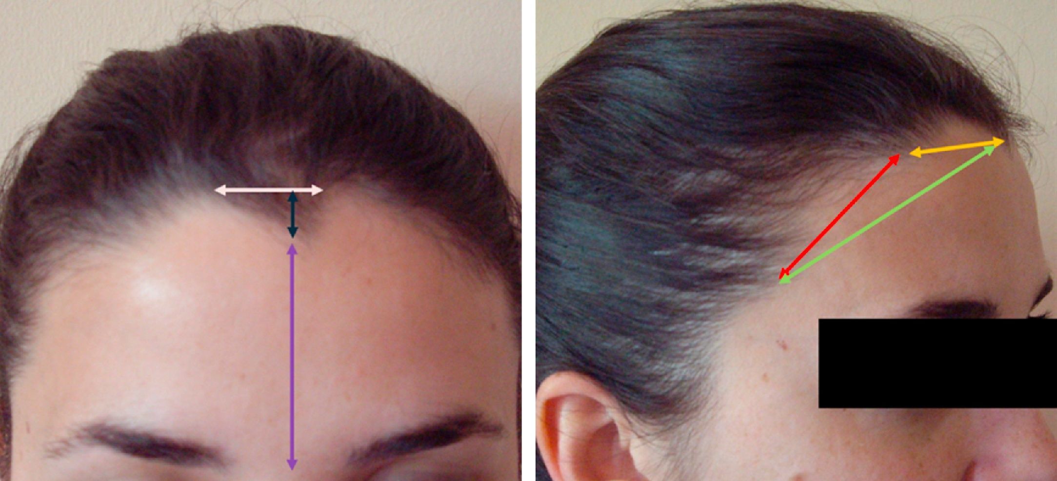
In a physical assessment, the presence and dimensions of the diverse structures that form the anterior hairline were measured and recorded as follows: a) widow's peak (V-shaped structure at the center of the anterior hairline), including the dimensions of the peak (width and height in centimeters) and the distance between the tip of the peak and the mid-eyebrow point; b) left and right lateral mounds, and in participants who had a widow's peak as well as lateral mounds (on one or both sides), we measured the distance between the apex of the widow's peak and that of each of the mounds; c) left and right temporal points; d) the distance between the widow's peak and the temporal points, whenever possible; and e) the distance between the lateral mound and the temporal point on both sides of the head (Fig. 1). All the measurements and photographs were obtained by the first 3 authors in accordance with the protocol established by the hospital's trichology unit.
- 5.
In a subgroup of 50 volunteers (98% physicians) who were not on contraceptives, the following hormonal, antigenic, and globulin profiles were assessed: follicle-stimulating hormone, luteinizing hormone, prolactin, 17-β estradiol, Δ-4-androstenedione, dehydroepiandrosterone sulfate, 17-hydroxyprogesterone, total and free testosterone, 5-α-dihydrotestosterone, prostate specific antigen, and sex hormone binding globulin. These tests were performed 3 to 5 days after the end of the menstrual cycle.
We recruited 103 healthy white Spanish women, of whom a subgroup of 50 participated in the hormone study. The mean age of the women was 29.72 years and the mean age at menarche was 12.34 years. Some 15.45% of the participants were smokers (mean number of cigarettes smoked per day, 10.39). The most prevalent characteristics of the group were skin phototype III (80.35%), brown hair (82.40%), and brown eyes (85.49%). Data on facial and body sebum and stratum corneum hydration for the 103 participants studied are summarized in Table 1.
A widow's peak was observed in 94.17% of the participants; this structure had a mean height of 1.01cm and a mean width of 2.13cm. Lateral mounds on both sides of the head were observed in all the women. The mean distances between the hairline structures are listed in Table 2.
Mean Distances Between Hairline Structures.
| Hairline Structures and Measurements | Mean, cm | Range, cm |
| Widow's peak: width | 2.13 | 0.00-5.00 |
| Widow's peak: height | 1.01 | 0.00-3.50 |
| Widow's peak to mid-eyebrow | 5.89 | 4.50-9.00 |
| Widow's peak to left lateral mound | 4.40 | 2.00-7.00 |
| Widow's peak to right lateral mound | 4.45 | 2.50-7.50 |
| Left lateral mound to left temporal point | 4.54 | 2.50-9.00 |
| Right lateral mound to right temporal point | 4.83 | 2.00-8.30 |
| Widow's peak to left temporal point | 8.15 | 5.00-10.00 |
| Widow's peak to right temporal point | 8.11 | 5.60-10.50 |
The results of the hormone studies for the 50 women who were not taking oral contraceptives are shown in Table 3; this subgroup had a mean age of 26.3 years.
Mean Hormone Levels and Normal Ranges.
| Hormones | Mean | Range | Reference Value |
| Follicle-stimulating hormone, mIU/mL | 5.59 | 2-9.2 | 4.00 to 13.00 |
| Luteinizing hormone, mIU/mL | 7.71 | 0.5-18.2 | 1.00 to 18.00 |
| Prolactin, μIU/mL | 216.01 | 46-501 | 70.00-600.00 |
| 17-β-estradiol, pg/mL | 93.80 | 7-455 | 12.00-170.00 |
| 17-OH-progesterone, ng/mL | 1.39 | 0.6-5.9 | 0.20-1.30 |
| 5α-dihydrotestosterone, pg/mL | 352.21 | 119-654 | 24.00-368.00 |
| Dehydroepiandrosterone sulfate, μg/dL | 175.24 | 41-390 | 80.00-400.00 |
| Total testosterone, nmol/L | 1.68 | 0.6-3 | 0.20-3.00 |
| Free testosterone, pg/mL | 1.12 | 0.13-4.43 | 0.00-4.50 |
| Δ4-androstenedione, ng/mL | 1.05 | 0.2-5.36 | 0.50-3.00 |
| Prostate specific antigen, ng/mL | 0.04 | 0.02-0.08 | 0.00-0.02 |
| Sex hormone binding globulin, nM/L | 95.33 | 2.44-162 | 20.00-130.00 |
No statistically significant associations between hormone levels in plasma and the height or width of the widow's peak or the other dimensions studied were detected (Spearman's ρ; P>.05).
DiscussionWith ever greater frequency, patients consulting their physician about hair and scalp health are interested in maintaining a healthy and youthful appearance. It may be possible to assist such patients by transplanting hair to achieve the appearance of greater density or to conceal areas of hair loss. It may even, occasionally, be possible to hide scars caused by scalp injury or surgery.
Transplanted hair follicles should be distributed in a way that imitates the pattern that is natural in the healthy woman in the interest of achieving satisfactory results.
While male hairline patterns were studied many years ago, the data for men cannot be extrapolated to women. In 2009, Nusbaum and Fuentefria10 published a pioneering study on natural female hairlines. While they determined the frequency, dimensions, and location of the structures that compose the hairline in 360 American female volunteers, they did not assess the clinical status of the participants (the study was carried out in hairdressing salons) or indicate whether the women were white, Hispanic, or of another ethnic group. The prevalence of the widow's peak was higher in our study (94% vs 81%). The widow's peaks in our sample also had greater height on average (1.01cm vs 0.83cm) and we measured greater distances between hairline structures: mean distance between the frontal midpoint and the mid-eyebrow, 5.89cm vs 5.5cm; mean distance between the frontal midpoint and the apex of the right lateral mound, 4.45cm vs 3.74cm; mean distance between the frontal midpoint and the apex of the left lateral mound, 4.40cm vs 3.97cm; and mean distance between the right lateral mound and the right temporal point, 4.83cm vs 3.78cm, and between the left lateral mound and the left temporal point, 4.54cm vs 3.51cm. These findings suggest that the contour of the anterior hairline in young, healthy white Spanish women tends to be more irregular than those previously characterized and that lateral mounds and widow's peaks are more pronounced. This difference could be racial in origin, but could also reflect the age difference between our participants (mean age, 29 years) and the group studied by Nusbaum and Fuentefria (mean age, 41 years), which included patients up to 70 years of age.
Jung et al.11 also described anterior hairlines in women, but they studied an Asian population and focused on the contour of the hairline, classifying it into 5 shapes or patterns: round, M-shaped, rectangular, triangular, and bell-shaped. However, they did not record the presence or dimensions of the typical hairline structures. Although it is interesting to have data on how the hairline frames the face, this information is of limited use to the surgeon considering surgical repair of a hairline because it cannot be compared to data from other studies Moreover, in some diseases, such as frontal fibrosing alopecia, the hairline may retain a normal contour but the distance from the mid-eyebrow point is altered. In our study, the contour can be extrapolated from the dimensions of the various structures that make up the hairline.
An interesting finding of the hormone study was the elevated mean concentration of 17-hydroxy-progesterone (1.39 ng/mL; range, 0.6-5.9 ng/mL), indicating the greater clearance of adrenal hormones characteristic of physical and intellectual activity. Also high considering that our participants were premenopausal was the mean concentration of prostate specific antigen (0.04 ng/mL; range, 0.02-0.08 ng/mL), a finding indicative of androgenetic activity.4,12 The relationship between all these markers requires more study along the same lines as ours but in other societal groups.
ConclusionsIn a series of healthy white Spanish women of childbearing age, we studied the frequency and size of the widow's peak, the dimensions of the structures that make up the anterior hairline, and the distances between these points. We also assessed sebum content and stratum corneum hydration on the forehead and arm. We believe that this data is useful for the assessment, grading, and treatment of hairline recession caused by conditions such as female androgenetic alopecia occurring in a male pattern and certain forms of scarring alopecia, such as frontal fibrosing alopecia. Likewise, we believe it will help practitioners to achieve more acceptable cosmetic results when surgical reconstruction of the hairline is undertaken because the patient wishes to have an anterior hairline similar to that of other white Spanish women.
Our data describe, for the first time, the shape and dimensions of the anterior hairline in young white Spanish women, a very homogeneous population in this respect. In the absence of broader population studies, this data can be used as a gold standard when surgical modification of the anterior hairline is required in a female patient. The differences between our findings and those of the few publications available may be related to differing racial and age profiles, and the possible presence of concomitant conditions in the American group. Similar studies are needed for other age groups in order to assess whether the hairline changes with age or with menopause, so that age-appropriate reconstruction can be achieved.
The relationship observed between the dimensions of the widow's peak, the mean sebum and hydration values, and the hormonal (17-hydroxy-progesterone) and antigenic (prostate specific antigen) markers, could be due to unforeseen bias introduced by recruiting a sample of white Spanish women who were mainly physicians (98%). The participants were therefore working in an occupation that involves a certain degree of emotional and intellectual stress, which can be associated with higher adrenal androgen clearance rates.
Further studies of these markers in other population groups would help detect the possible influence of hormonal changes on the distribution of the anterior hairline, sebum secretion, and stratum corneum hydration.
Ethical DisclosuresProtection of human and animal subjectsThe authors declare that no experiments were performed on humans or animals for this investigation.
Confidentiality of dataThe authors declare that this study was carried out in accordance with the protocols of their institution concerning the publication of patient data, and that all the participants included in the study were properly informed and gave their written informed consent to participation.
Right to privacy and informed consentThe authors obtained the informed consent of the patients and/or subjects referred to in this article. The signed forms are in the possession of the corresponding author.
Conflicts of InterestThe authors declare that they have no conflicts of interests.
Please cite this article as: Ceballos C, et al. Estudio de patrones de implantación pilosa frontal en la mujer española caucásica. Actas Dermosifiliogr. 2013;104:311–5.


