

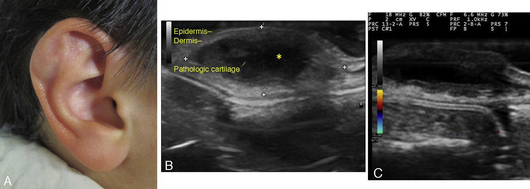
A 9-year-old boy was seen for an asymptomatic lesion of 7 months duration on the right pinna. The lesion first appeared after trauma and had grown progressively since then. The lesion had been drained twice with subsequent recurrence. Culture of the serous effusion was negative. The physical examination showed an indurated fluctuant nodule measuring 1.6 by 0.9cm on the superior right antihelix (Fig. 1a). Skin ultrasound revealed a hypoechoic cystic lesion with anechoic content, no wall (asterisk), and posterior reinforcement, measuring 2.09 by 1.58cm (Fig. 1b; longitudinal section, B mode, 18Mhz). There was no increased intralesional or perilesional blood flow in the Doppler images (Fig. 1c). The lesion was diagnosed as an auricular pseudocyst and the patient was referred for pediatric surgery. Auricular pseudocyst is an uncommon entity that presents as an asymptomatic nodule. It consists of a cystic formation whose wall is formed of partially degenerative auricular cartilage. The etiology of lesion is not clear, but trauma to an exposed area is the most widely accepted possibility. The lesion is usually treated with surgery or by corticosteroid infiltration. Recurrences are common. Use of skin ultrasound in such lesions has very rarely been reported. In our case, this technique was very useful for confirming diagnosis. In addition, the technique could be of use for early detection of recurrence and for guiding infiltration of corticosteroids or other drugs.
Please cite this article as: Blasco-Morente G, Arias-Santiago S, Garrido-Colmenero C, Pérez-López I. Características ecográficas del psudoquiste auricular. Actas Dermosifiliogr. 2015;106:840.


