


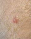
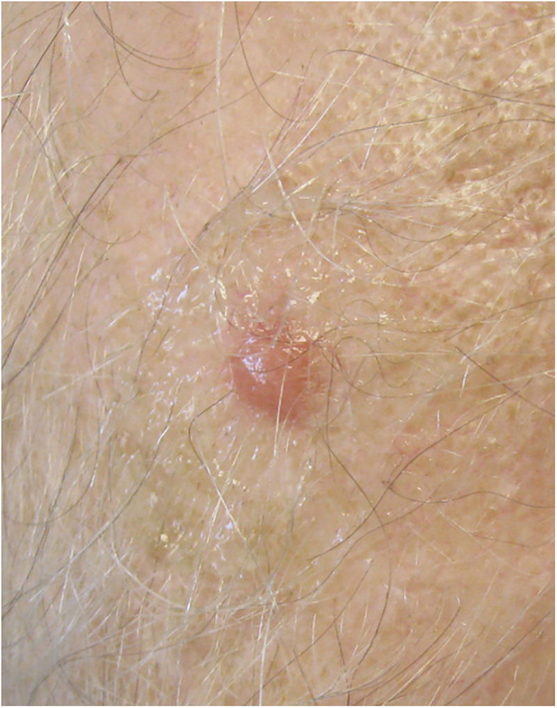
A 66-year-old man presented with orange, noninfiltrated, fibrous papulonodular lesions that had grown progressively on the head, neck, and upper trunk (Fig. 1).
What Is Your Diagnosis?
Cutaneous metastasis of pancreatic adenocarcinoma.
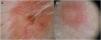
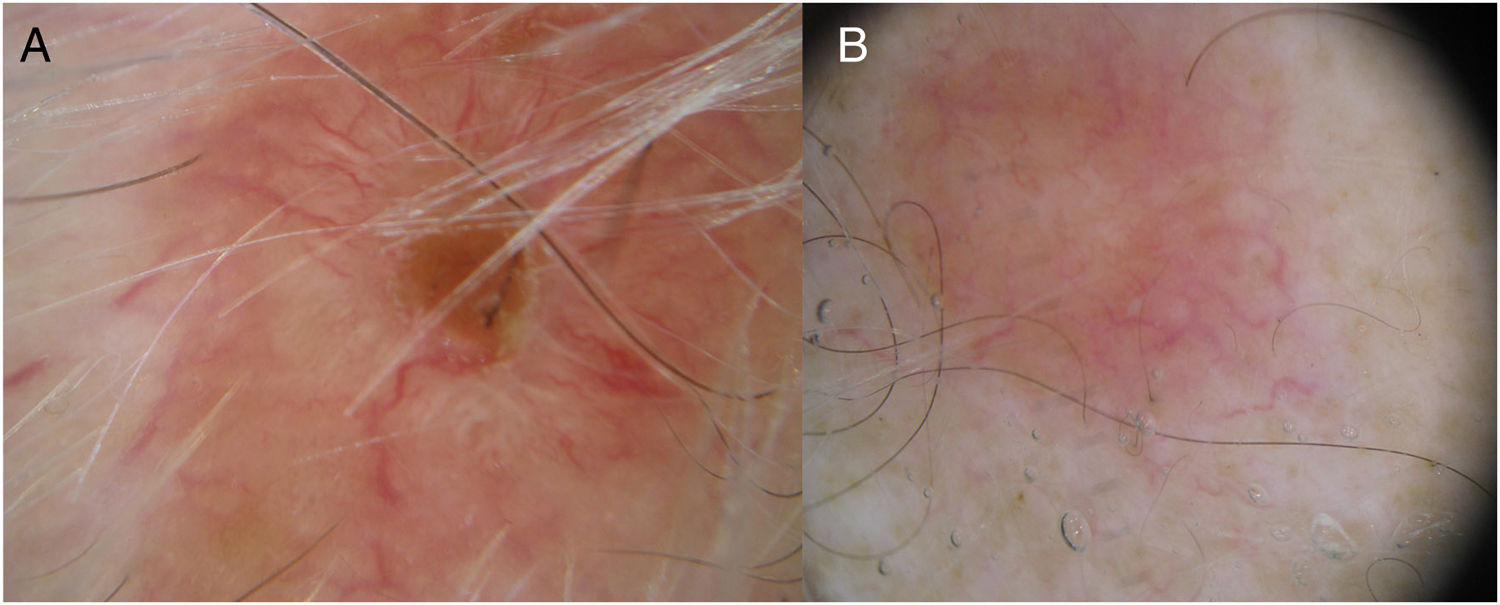
CommentDermoscopy showed an orange background and centripetally arranged, tortuous, branched vascularization (Fig. 2). Analysis of one of the lesions revealed dermal proliferation of epithelial cells, which were either isolated or formed small nests and trabeculae. Immunohistochemistry revealed positive staining for CK7 and negative staining for CK20 and CDX2. Taken together, the findings were compatible with cutaneous metastasis of pancreatic adenocarcinoma.
An orange background on dermoscopy should raise suspicion of histiocytic aggregates, a finding associated with a broad spectrum of lesions, including xanthoma, xanthogranuloma, sarcoidosis, lupus vulgaris, and leishmaniasis. Less frequently, this finding is found in other lesions, such as cylindroma.1
Cutaneous metastasis is a relatively infrequent finding (0.6–9% of tumors), the presence of which primarily depends on the stage and histological subtype of the primary tumor. The clinical presentation is highly variable. In some cases cutaneous metastasis can mimic angiomas or cysts, among other entities.2,3 The most common dermoscopic finding (in up to 88% of cases) is the presence of vascular structures,2 which are predominantly serpentine, irregular, punctate, corkscrew-like, or branched, are usually small in caliber, and may disappear when pressed.2–4 In a series published by Chernoff and coworkers,2 the most frequent finding was serpentine vascularization, followed by arboriform vascularization, although irregular polymorphic vascularization, as described in our patient, was also very frequent (59% of cases).
Dermoscopy of cutaneous metastasis generally reveals a pink background and an absence of structures,2–4 although pigmented areas are observed in some cases. These pigmented structures may have a melanocytic pattern (e.g. brownish lines or blue-gray blood cells),2 as described in metastatic breast carcinoma.2,5 Cutaneous metastasis can also mimic other pigmented lesions, including dermatofibroma.6
In conclusion, we describe an atypical case of cutaneous metastasis of pancreatic cancer characterized by an orange background, a finding not previously associated with this type of lesion.
Conflicts of InterestThe authors declare that they have no conflicts of interest.
Please cite this article as: Gonazlez-Delgado VA, Cordero-Romero P, Martín JM. Lesiones anaranjadas de aparición progresiva en cuero cabelludo. Actas Dermosifiliogr. 2020;111:317–318.


