

Actinic keratosis is a skin disease with the potential to progress to cutaneous squamous cell carcinoma, making its treatment essential. However, the last update of the Spanish adaptation of the European clinical guidelines dates back to 2014. This document includes the recommendations agreed upon by 75 primary care and dermatology experts on the optimal management of patients with actinic keratosis. In general, early and detailed diagnosis of actinic keratosis using dermoscopy and referral to dermatology is recommended, especially in high-risk patients. Regarding treatment, experts recommend the use of treatments adapted to the degree and extent of the injuries, highlighting the use of molecules such as 5-fluorouracil for isolated and field of cancerization injuries, as well as tirbanibulin for grade 1 and 2 isolated and field of cancerization injuries. These consensual recommendations seek to serve as a clinical guide on the routine management of patients with actinic keratosis.
Actinic keratosis (AK) is a chronic dermatologic disease caused by the intradermal proliferation of atypical keratinocytes following prolonged exposure to ultraviolet radiation.1 Patients present with erythematous lesions of variable color—ranging from light to reddish or pigmented—and with a rough texture, typically located in sun-exposed areas such as the face, neck, and dorsal hands.2 The prevalence of AK in Spain is difficult to determine due to regional variability,3 but it is estimated to be above 15%, similar to other European regions.4
AK is more common in men, older individuals, those with fair skin, and those with a history of chronic sun exposure.5 Importantly, affected cells have the potential to transform into cutaneous squamous cell carcinoma (cSCC), the 2nd most common skin cancer worldwide.6 This risk varies according to time, number of lesions, and the patient's immune status.7 However, the absence of treatment leads to progression to cSCC in approximately 20% of cases, and 60% of cSCCs arise in areas affected by AK.8,9 Diagnosis is based on clinical evaluation, sometimes along with histopathological analysis to differentiate AK from other lesions such as invasive cSCC, and to distinguish pigmented AK from lentigo maligna.10,11 Although this diagnosis can be performed in primary care, in Spain it is predominantly established in specialized dermatology settings,3–12 accounting for 28.6% of dermatologic consultations.13 In terms of pathology, patients may present with isolated lesions or field cancerization. AK is categorized into 3 grades of severity: grade 1 and grade 2 denote atypical keratinocytes occupying the lower third and the lower two-thirds of the epidermis, respectively; grade 3 refers to thick hyperkeratotic plaques extending beyond two-thirds of the epidermis, associated with a higher probability of progression to cSCC. Nonetheless, all AKs, from grade 1 to grade 3, have the potential to evolve into cSCC.2–14
The primary endpoint of AK treatment is to eliminate clinical lesions to prevent progression to cSCC and to reduce the formation of new lesions within the field cancerization area.15,16 When lesions are few in number and/or isolated, ablative treatments are typically used, with cryosurgery or cryotherapy being more frequent than curettage combined with electrocoagulation. Conversely, in patients with extensive involvement, dynamic photodynamic therapy and topical treatments—such as 5-fluorouracil (5-FU), imiquimod, diclofenac, or tirbanibulin—are recommended.17,18 However, treatment success depends on individual patient characteristics and on the chronic nature of the disease, which necessitates long-term therapies that may reduce adherence and complicate AK control.18
The most recent Spanish adaptation of the European guideline for the management of AK dates back to 2014.19 More recently, a multidisciplinary expert consensus from several European dermatology associations issued updated recommendations for the diagnosis and treatment of AK. Among these, individualized strategies are strongly emphasized, especially for patients at higher risk of developing cSCC, such as immunosuppressed individuals.20 For this reason, the present study aimed to update diagnostic and therapeutic recommendations for the optimal management of patients with AK.
Materials and methodsThe study was conducted in several phases: literature review, focus group, and Delphi consultation.
Literature reviewWe conducted an organized review of the literature to identify available evidence regarding AK management using the Medline/PubMed international database. Searches employed filters and Medical Subject Headings (MeSH) combined with open search terms and Boolean connectors “OR” and “AND.” Additionally, a structured manual search of gray literature was performed on the websites of leading scientific societies such as the Spanish Academy of Dermatology and Venereology (AEDV), the Spanish Society of Primary Care Physicians (SEMERGEN), and the European Association of Dermato-Oncology, among others. Articles published in English or Spanish within the last 10 years (2014–2024) were included. Exclusion criteria were commentaries, letters to the editor, editorials, book chapters, and publications unrelated to AK.
Focus groupA total of 6 experts in AK management (3 in dermatology and 3 in primary care) formed the scientific committee and focus group. With the support of a moderator, the group reviewed the collected evidence to develop the questionnaire used in the Delphi consultation. The focus group also formulated the final list of recommendations based on the consensus statements.
Delphi consultationQuestionnaireThe questionnaire comprised 4 sections: (1) sociodemographic and professional characteristics (6 items); (2) diagnosis of AK (21 statements); (3) referral criteria and pathways (13 statements); and (4) therapeutic management (66 statements).
Consultation and panelistsThe Delphi consultation was directed at primary care physicians and dermatologists with at least 2 years of experience managing AK within the public Spanish National Health System. The consultation was conducted over 2 consecutive rounds, where panelists rated their level of agreement with each statement using a 7-point Likert scale (1=strongly disagree; 7=strongly agree). A consensus threshold of 70% agreement (Likert 6–7) or disagreement (Likert 1–2) was established. Statements not reaching consensus in the first round were reassessed in the second round.
Panelists were identified and invited to participate by SEMERGEN and AEDV, and those who agreed received the questionnaire electronically.
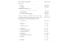
ResultsA total of 75 experts completed the 1st round and 68 completed the 2nd (90.7% response rate). Their characteristics are detailed in Table 1.
Sociodemographic and professional characteristics of the panelists.
| Age in years; mean (SD) | 44.9 (10.2) |
| Sex; n (%) | |
| • Male | 28 (37.3) |
| • Female | 47 (62.7) |
| Professional profile; n (%) | |
| • Family and Community Medicine | 16 (21.3) |
| • Dermatology | 30 (40.0) |
| • Oncologic Dermatology | 29 (38.7) |
| Years of experience managing AK; mean (SD) | 16.8 (9.6) |
| Number of patients seen per month; mean (SD) | 90.1 (91.6) |
| Autonomous community of practice; n (%) | |
| • Andalusia | 9 (12.0) |
| • Aragón | 6 (8.0) |
| • Asturias | 1 (1.3) |
| • Balearic Islands | 2 (2.7) |
| • Canary Islands | 4 (5.3) |
| • Cantabria | 1 (1.3) |
| • Castile-La Mancha | 3 (4.0) |
| • Castile and León | 6 (8.0) |
| • Catalonia | 14 (18.7) |
| • Valencian Community | 6 (8.0) |
| • Galicia | 1 (1.3) |
| • Madrid | 21 (28.0) |
| • Navarre | 1 (1.3) |
SD, standard deviation; AK, actinic keratosis.
In the Delphi survey, 70 of the 100 statements presented (70%) reached consensus. All statements related to the diagnosis of AK (21) and the referral criteria and pathways (13) achieved consensus (Supplementary Tables 1 and 2). Similarly, of the 66 statements proposed regarding therapeutic management, 36 (54.5%) reached consensus (including 4 statements with consensus in disagreement) (Supplementary Tables 3–8). The final list of recommendations is shown in Table 2.
List of recommendations.
| Diagnosis |
| Whenever possible, the use of dermatoscopes in primary care is recommended to improve diagnostic accuracy and to provide relevant information for appropriate referral to dermatology, particularly when referral occurs via teleconsultation.In general, the diagnosis of AK should be made through physical examination, with histological diagnosis recommended only in: (1) cases with unclear clinical findings; (2) presence of clinical signs of progression to cutaneous squamous cell carcinoma; and/or (3) resistance to therapy.During diagnosis, it is recommended to evaluate modulating risk factors (age, skin phototype, baldness, lifestyle, occupation, geographic location, history of skin cancer or immunosuppression) and to document the location, number, and severity of AK lesions.The following clinical signs indicate possible progression of AK to cutaneous squamous cell carcinoma: (1) thickening or induration; (2) pain on palpation, bleeding, or ulceration; (3) rapid growth; (4) surface changes; (5) resistance to therapy; and/or (6) rapid recurrence after initially successful treatment.A patient is considered to have a field of cancerization when at least 6 AK lesions are present in a body region or field, with evidence of actinic skin damage and hyperkeratosis in contiguous areas.The use of the “5 Rs+R rule” (Red, Rough, Recurrent lesions in sun-exposed Regions receiving Radiation+Risk of malignant transformation) is recommended for AK diagnosis, especially in primary care.Training on the diagnosis and management of AK—particularly directed at primary care professionals—is recommended. Whenever possible, the use of teledermatology is recommended to ensure accurate and timely diagnosis and follow-up. |
| Referral criteria |
| For appropriate referral of a patient with AK via teleconsultation, it is recommended—whenever feasible—to include a clinical photograph, a dermoscopic image, prior treatments, and risk factors in the health record. Referral of a patient with AK to dermatology is recommended in the following situations:– Immunosuppressed patients and solid-organ transplant recipients– Patients with clinical signs of possible progression to cutaneous squamous cell carcinoma– Patients with marked evidence of sun damage– Diagnosis of xeroderma pigmentosum– Multiple AK lesions in highly susceptible areas such as the face, scalp, or hands– AK lesions located in high-risk regions (periocular area, ears, lips)– Uncertain diagnosis requiring specialist confirmation– Treatment failure– Multiple or recurrent AK lesions difficult to treat |
| Therapeutic management |
| The use of the following treatments for managing AK lesions is recommended (green) or discouraged (red) depending on lesion type and grade. |
| Lesion grade | Isolated lesions | Lesions with field cancerization |
|---|---|---|
| Grade 1–2 | ✓ Cryosurgery✓ 4% 5-fluorouracil✓ 0.5% 5-fluorouracil with 10% salicylic acid✓ 1% tirbanibulin | ✓ 4% 5-fluorouracil✓ 5% imiquimod✓ 3.75% imiquimod✓ 3% diclofenac sodium with 2.5% hyaluronic acid✓ 1% tirbanibulin✓ Conventional photodynamic therapy✓ Daylight photodynamic therapy× Curettage/electrocoagulation |
| Grade 3 | ✓ Cryosurgery✓ Curettage/electrocoagulation✓ 0.5% 5-fluorouracil with 10% salicylic acid | ✓ 4% 5-fluorouracil✓ 5% imiquimod✓ 3.75% imiquimod✓ Conventional photodynamic therapy× Curettage/electrocoagulation× 3% diclofenac sodium with 2.5% hyaluronic acid |
| In the case of combination treatments, the following therapies are recommended for both isolated lesions and lesions with field cancerization:5-FU followed by cryosurgery, cryosurgery followed by imiquimod, or cryosurgery followed by 5-FU. | ||
| In immunodeficient and immunosuppressed patients with AK lesions,cryosurgery, curettage, 5-fluorouracil (4% and 0.5%), and photodynamic therapy—both conventional and daylight—are generally recommended. | ||
| When selecting the most appropriate treatment for AK lesions,the patient's ability to self-administer therapy or the availability of caregivers/family members to do so should be taken into consideration. | ||
| After diagnosing AK,patients should be encouraged to adopt photoprotection measures, including behavioral changes regarding sun exposure, the use of sunscreen and protective clothing, as well as promoting self-examination. | ||
| Patients with AK should be followed periodically,with follow-up intervals adjusted according to the number of lesions, patient profile, and associated risk factors, particularly in immunodeficient and immunosuppressed patients. | ||
| Educational and awareness campaigns are recommendedto improve knowledge and prevention of AK among patients, families, and caregivers. | ||
5-FU, 5-fluorouracil; AK, actinic keratosis.
Spanish and European clinical practice guidelines emphasize the need to perform histopathological analyses when diagnosing lesions suspected of progressing to cutaneous squamous cell carcinoma (cSCC).19,20 This recommendation is particularly relevant in cases of actinic cheilitis, which has a malignant transformation rate between 10% and 30% and accounts for 95% of cSCCs arising on the lips.21 The panelists reached consensus on these aspects, including the management of actinic cheilitis—an aspect not addressed in the latest Spanish adaptation of the European clinical practice guidelines on the management of AK.19
In addition, the panelists recommended collecting sociodemographic information and the patient's health history. This aligns with findings from the Rotterdam Study, in which male sex, age older than 70 years, fair skin phototype, a prior history of skin cancer, and residence in regions with high sun exposure were identified as significant risk factors for AK.5
Consistent with these risk factors, it is essential to consider associated symptoms and their clinical implications. A prospective study demonstrated that manifestations such as pruritus, pain, bleeding, or changes in lesion size occur in 20–50% of patients with cSCC.11 In this context, panelists recommend assessing these signs and symptoms using the “5R+R” methodology proposed by Domínguez-Cruz et al., which facilitates a simple and systematic approach to diagnosing AK and detecting potential progression to cSCC.22 Similarly, to identify the presence of field cancerization, the panelists advise evaluating the number of lesions and the adjacent photodamaged skin, which is consistent with a previous consensus that highlighted the ambiguity surrounding the definition of field cancerization in the literature and advocated for the use of anatomical indicators as evaluation criteria.23
Complementing these considerations, diagnostic support tools play a crucial role. In line with the conclusions of the above-mentioned consensus (Figueras Nart et al.), the panelists recommend the use of dermoscopy in primary care, as it facilitates distinguishing AK from other conditions such as superficial basal cell carcinoma.19 Combining dermoscopy with appropriate training optimizes clinical diagnosis and helps determine the need for early referral to dermatology.24,25 However, widespread dermoscopy use still faces limitations, such as insufficient equipment availability.26
The use of telemedicine tools has expanded exponentially, especially in dermatology, due to the increasing number of consultations and advances in digital imaging.27 Teledermoscopy is widely recommended because it significantly improves diagnostic accuracy (92.4% with teledermoscopy vs 62.4% without it in primary care; p<0.001),28 thereby accelerating diagnostic pathways.29 Experts recommend providing all relevant information to ensure appropriate patient referral—particularly in teleconsultations—such as including a dermoscopic image in addition to a clinical photograph and the patient's health history.29,30
Recommendations on referral criteria and pathwaysPanelists recommend that the patient's health record should include a clinical photograph, a dermoscopic photograph, previous treatments, and risk factors.2,30 This recommendation aligns with prior dermatology guidelines and National Health Service (UK) documents.16,31 The authors support this suggestion given existing evidence of a lack of standardized referral criteria for AK patients from primary care to dermatology.26,29,32 In line with these guidelines, panelists advise referral especially for patients with risk factors such as xeroderma pigmentosum, a history of extensive sun damage, or lesions in anatomically complex areas (periocular region, lips)16,31–33—regions that not only have higher malignant potential but also pose therapeutic challenges.16,31,33
Recommendations on therapeutic managementBecause AK is considered a carcinoma in situ with the potential to progress to cSCC, previous guidelines recommend treating all AKs, including early or incipient lesions.14,19 In this regard, guidelines state that treatment of choice should be based on factors such as lesion extent and severity,19,31 recommending cryotherapy for isolated lesions, especially in patients with <6 lesions or unresponsive to topical therapies.15 A systematic review reported AK clearance rates >70% within 1–12 months after cryotherapy, with increased efficacy when combined with 0.5% 5-fluorouracil (5-FU), resulting in a mean improvement of 13.3%.18 Additionally, a clinical trial showed that cryotherapy followed by imiquimod significantly reduced lesion numbers vs control (78 vs. 116).34 For these reasons, both combinations were recommended by the panelists.
Curettage is not only effective for treating isolated grade 3 lesions but also enables histopathologic sampling.35 However, panelists discouraged its use for extensive involvement, as it is a painful procedure requiring local anesthesia15,36 and because clinical trial evidence is limited.
Regarding topical therapies, panelists recommended 5-FU for both isolated lesions (0.5% with 10% salicylic acid) and for field cancerization (4%). A meta-analysis showed lesion reductions up to 80.1% at 3 months and 67.4% at 6 months, although efficacy is strongly influenced by treatment adherence, which may be affected by adverse effects such as pruritus and burning sensation.37 Notably, combining 0.5% 5-FU with 10% salicylic acid improves penetration and enhances therapeutic action.38
Imiquimod, at both 3.75% and 5%, was recommended for field cancerization regardless of severity. Reviews agree that formulation and frequency should be tailored to each patient.15 The 5% formulation has demonstrated lesion clearance rates of up to 85%,37 whereas the 3.75% formulation shows similar efficacy with fewer adverse events.39
Regarding diclofenac sodium, panelists recommended its use for grade 1–2 lesions, both isolated and in field cancerization, highlighting that combining it with hyaluronic acid enhances drug penetration.40 However, its use was discouraged for grade 3 lesions due to its lower efficacy (36% lesion reduction) vs cryotherapy (72.3%) and 5-FU (80.1%).18 For tirbanibulin, clinical trials have demonstrated that 1% formulations achieve complete clearance in approximately half of patients with mild to moderate involvement and have a favorable safety profile.41 Accordingly, panelists recommended its use for grade 1–2 lesions, both isolated and in field cancerization.
Consistent with a recent narrative review,42 panelists recommended photodynamic therapy (PDT) for patients with field cancerization. While daylight-PDT has been described as less painful,43 a recent meta-analysis found that although efficacy is similar to conventional PDT for isolated grade 1–2 lesions (RR, 0.97; 95%CI, 0.91–1.04; p=0.41), it is less effective for grade 3 lesions (RR, 0.87; 95%CI, 0.81–9.94; p<0.001).43
Similarly, for immunosuppressed patients, panelists advised using conventional therapies such as cryotherapy, curettage, 5-FU, and both conventional and daylight-PDT, given the reduced efficacy of alternative therapies, the higher risk of cSCC, and the lack of clinical trials evaluating newer therapies in this population.44,45
Finally, panelists emphasized the importance of incorporating the patient's perspective into treatment decisions, as motivation and engagement strongly influence adherence.46 A cross-sectional study showed that patients prioritize preventing AK progression to cSCC but also value cosmetic outcomes and treatment convenience—especially in older adults.47 In line with these observations, experts recommend patient education on self-examination and treatment administration, along with periodic follow-up, which may improve adherence and optimize therapeutic outcomes.
ConclusionsPanelists highlighted the importance of improving diagnostic processes in primary care to optimize appropriate referrals to dermatology. For treatment, they agreed on using tailored strategies based on patient profile and lesion type, emphasizing 0.5% 5-FU for isolated lesions and 4% 5-FU for field cancerization, as well as tirbanibulin for grade 1 and 2 lesions, either isolated or in field cancerization. Finally, experts stressed the importance of patient education to optimize treatment adherence. This document may serve as a clinical guide to facilitate diagnosis, referral, and treatment of patients with AK in routine clinical practice. Interpretation of the findings should consider the inherent limitations of this study and its context within the Spanish health care system.
FundingThis study was funded by Almirall as part of an agreement to promote independent research. The funding entity had no role in the design, conduct, analysis, or interpretation of the study results.
The authors would like to thank all the professionals who participated in the Delphi consultation for their valuable contribution to the development of this study. They also thank Outcomes’10 for providing methodological support and manuscript writing services.
The followings are the supplementary data to this article:


