array:24 [
  "pii" => "S1578219018304165"
  "issn" => "15782190"
  "doi" => "10.1016/j.adengl.2018.11.018"
  "estado" => "S300"
  "fechaPublicacion" => "2019-01-01"
  "aid" => "1901"
  "copyright" => "Elsevier España, S.L.U. and AEDV"
  "copyrightAnyo" => "2018"
  "documento" => "simple-article"
  "crossmark" => 1
  "subdocumento" => "crp"
  "cita" => "Actas Dermosifiliogr. 2019;110:71-4"
  "abierto" => array:3 [
    "ES" => true
    "ES2" => true
    "LATM" => true
  ]
  "gratuito" => true
  "lecturas" => array:2 [
    "total" => 3
    "HTML" => 3
  ]
  "Traduccion" => array:1 [
    "es" => array:19 [
      "pii" => "S0001731018300553"
      "issn" => "00017310"
      "doi" => "10.1016/j.ad.2017.11.012"
      "estado" => "S300"
      "fechaPublicacion" => "2019-01-01"
      "aid" => "1901"
      "copyright" => "AEDV"
      "documento" => "simple-article"
      "crossmark" => 1
      "subdocumento" => "crp"
      "cita" => "Actas Dermosifiliogr. 2019;110:71-4"
      "abierto" => array:3 [
        "ES" => true
        "ES2" => true
        "LATM" => true
      ]
      "gratuito" => true
      "lecturas" => array:2 [
        "total" => 249
        "formatos" => array:2 [
          "HTML" => 111
          "PDF" => 138
        ]
      ]
      "es" => array:11 [
        "idiomaDefecto" => true
        "cabecera" => "<span class="elsevierStyleTextfn">CARTA CIENT&#205;FICO-CL&#205;NICA</span>"
        "titulo" => "Milia&#44; una reacci&#243;n infrecuente tras terapia fotodin&#225;mica"
        "tienePdf" => "es"
        "tieneTextoCompleto" => "es"
        "paginas" => array:1 [
          0 => array:2 [
            "paginaInicial" => "71"
            "paginaFinal" => "74"
          ]
        ]
        "titulosAlternativos" => array:1 [
          "en" => array:1 [
            "titulo" => "Milia&#58; An Uncommon Reaction to Photodynamic Therapy"
          ]
        ]
        "contieneTextoCompleto" => array:1 [
          "es" => true
        ]
        "contienePdf" => array:1 [
          "es" => true
        ]
        "resumenGrafico" => array:2 [
          "original" => 0
          "multimedia" => array:7 [
            "identificador" => "fig0010"
            "etiqueta" => "Figura 2"
            "tipo" => "MULTIMEDIAFIGURA"
            "mostrarFloat" => true
            "mostrarDisplay" => false
            "figura" => array:1 [
              0 => array:4 [
                "imagen" => "gr2.jpeg"
                "Alto" => 975
                "Ancho" => 1300
                "Tamanyo" => 369300
              ]
            ]
            "descripcion" => array:1 [
              "es" => "<p id="spar0010" class="elsevierStyleSimplePara elsevierViewall">Estructura qu&#237;stica revestida de una pared de epitelio escamoso con capa granulosa y queratina ortoquerat&#243;sica en su interior&#46;</p>"
            ]
          ]
        ]
        "autores" => array:1 [
          0 => array:2 [
            "autoresLista" => "V&#46; Flores-Climente, J&#46; Dalmau, L&#46; Puig"
            "autores" => array:3 [
              0 => array:2 [
                "nombre" => "V&#46;"
                "apellidos" => "Flores-Climente"
              ]
              1 => array:2 [
                "nombre" => "J&#46;"
                "apellidos" => "Dalmau"
              ]
              2 => array:2 [
                "nombre" => "L&#46;"
                "apellidos" => "Puig"
              ]
            ]
          ]
        ]
      ]
      "idiomaDefecto" => "es"
      "Traduccion" => array:1 [
        "en" => array:9 [
          "pii" => "S1578219018304165"
          "doi" => "10.1016/j.adengl.2018.11.018"
          "estado" => "S300"
          "subdocumento" => ""
          "abierto" => array:3 [
            "ES" => true
            "ES2" => true
            "LATM" => true
          ]
          "gratuito" => true
          "lecturas" => array:1 [
            "total" => 0
          ]
          "idiomaDefecto" => "en"
          "EPUB" => "https://multimedia.elsevier.es/PublicationsMultimediaV1/item/epub/S1578219018304165?idApp=UINPBA000044"
        ]
      ]
      "EPUB" => "https://multimedia.elsevier.es/PublicationsMultimediaV1/item/epub/S0001731018300553?idApp=UINPBA000044"
      "url" => "/00017310/0000011000000001/v1_201901030610/S0001731018300553/v1_201901030610/es/main.assets"
    ]
  ]
  "itemSiguiente" => array:19 [
    "pii" => "S1578219018304086"
    "issn" => "15782190"
    "doi" => "10.1016/j.adengl.2018.11.010"
    "estado" => "S300"
    "fechaPublicacion" => "2019-01-01"
    "aid" => "1915"
    "copyright" => "Elsevier Espa&#241;a&#44; S&#46;L&#46;U&#46; and AEDV"
    "documento" => "simple-article"
    "crossmark" => 1
    "subdocumento" => "crp"
    "cita" => "Actas Dermosifiliogr. 2019;110:74-5"
    "abierto" => array:3 [
      "ES" => true
      "ES2" => true
      "LATM" => true
    ]
    "gratuito" => true
    "lecturas" => array:1 [
      "total" => 0
    ]
    "en" => array:11 [
      "idiomaDefecto" => true
      "cabecera" => "<span class="elsevierStyleTextfn">Case and Research Letters</span>"
      "titulo" => "Treatment of a Refractory Venous Ulcer With Terminal Interruption of the Reflux Source"
      "tienePdf" => "en"
      "tieneTextoCompleto" => "en"
      "paginas" => array:1 [
        0 => array:2 [
          "paginaInicial" => "74"
          "paginaFinal" => "75"
        ]
      ]
      "titulosAlternativos" => array:1 [
        "es" => array:1 [
          "titulo" => "Tratamiento de &#250;lcera venosa refractaria mediante la &#171;interrupci&#243;n terminal de la fuente de reflujo&#187;"
        ]
      ]
      "contieneTextoCompleto" => array:1 [
        "en" => true
      ]
      "contienePdf" => array:1 [
        "en" => true
      ]
      "resumenGrafico" => array:2 [
        "original" => 0
        "multimedia" => array:7 [
          "identificador" => "fig0005"
          "etiqueta" => "Figure 1"
          "tipo" => "MULTIMEDIAFIGURA"
          "mostrarFloat" => true
          "mostrarDisplay" => false
          "figura" => array:1 [
            0 => array:4 [
              "imagen" => "gr1.jpeg"
              "Alto" => 878
              "Ancho" => 1309
              "Tamanyo" => 192054
            ]
          ]
          "descripcion" => array:1 [
            "en" => "<p id="spar0005" class="elsevierStyleSimplePara elsevierViewall">A&#44; Venous ulcer before treatment&#46; B&#44; Complete resolution 4 weeks after beginning treatment&#44; with evident improvement of the surrounding skin&#46;</p>"
          ]
        ]
      ]
      "autores" => array:1 [
        0 => array:2 [
          "autoresLista" => "M&#46; Mendieta-Eckert, J&#46;L&#46; Azpiazu-Macho, N&#46; Landa-Gundin"
          "autores" => array:3 [
            0 => array:2 [
              "nombre" => "M&#46;"
              "apellidos" => "Mendieta-Eckert"
            ]
            1 => array:2 [
              "nombre" => "J&#46;L&#46;"
              "apellidos" => "Azpiazu-Macho"
            ]
            2 => array:2 [
              "nombre" => "N&#46;"
              "apellidos" => "Landa-Gundin"
            ]
          ]
        ]
      ]
    ]
    "idiomaDefecto" => "en"
    "Traduccion" => array:1 [
      "es" => array:9 [
        "pii" => "S0001731018300772"
        "doi" => "10.1016/j.ad.2017.10.024"
        "estado" => "S300"
        "subdocumento" => ""
        "abierto" => array:3 [
          "ES" => true
          "ES2" => true
          "LATM" => true
        ]
        "gratuito" => true
        "lecturas" => array:1 [
          "total" => 0
        ]
        "idiomaDefecto" => "es"
        "EPUB" => "https://multimedia.elsevier.es/PublicationsMultimediaV1/item/epub/S0001731018300772?idApp=UINPBA000044"
      ]
    ]
    "EPUB" => "https://multimedia.elsevier.es/PublicationsMultimediaV1/item/epub/S1578219018304086?idApp=UINPBA000044"
    "url" => "/15782190/0000011000000001/v1_201901030631/S1578219018304086/v1_201901030631/en/main.assets"
  ]
  "itemAnterior" => array:19 [
    "pii" => "S1578219018304177"
    "issn" => "15782190"
    "doi" => "10.1016/j.adengl.2017.09.007"
    "estado" => "S300"
    "fechaPublicacion" => "2019-01-01"
    "aid" => "1902"
    "copyright" => "Elsevier Espa&#241;a&#44; S&#46;L&#46;U&#46; and AEDV"
    "documento" => "simple-article"
    "crossmark" => 1
    "subdocumento" => "crp"
    "cita" => "Actas Dermosifiliogr. 2019;110:69-71"
    "abierto" => array:3 [
      "ES" => true
      "ES2" => true
      "LATM" => true
    ]
    "gratuito" => true
    "lecturas" => array:2 [
      "total" => 5
      "formatos" => array:2 [
        "HTML" => 2
        "PDF" => 3
      ]
    ]
    "en" => array:11 [
      "idiomaDefecto" => true
      "cabecera" => "<span class="elsevierStyleTextfn">Case and Research Letters</span>"
      "titulo" => "Extragenital Lichen Sclerosus Induced by Radiotherapy"
      "tienePdf" => "en"
      "tieneTextoCompleto" => "en"
      "paginas" => array:1 [
        0 => array:2 [
          "paginaInicial" => "69"
          "paginaFinal" => "71"
        ]
      ]
      "titulosAlternativos" => array:1 [
        "es" => array:1 [
          "titulo" => "Liquen escleroso extragenital inducido por radioterapia"
        ]
      ]
      "contieneTextoCompleto" => array:1 [
        "en" => true
      ]
      "contienePdf" => array:1 [
        "en" => true
      ]
      "resumenGrafico" => array:2 [
        "original" => 0
        "multimedia" => array:7 [
          "identificador" => "fig0005"
          "etiqueta" => "Figure 1"
          "tipo" => "MULTIMEDIAFIGURA"
          "mostrarFloat" => true
          "mostrarDisplay" => false
          "figura" => array:1 [
            0 => array:4 [
              "imagen" => "gr1.jpeg"
              "Alto" => 1005
              "Ancho" => 2000
              "Tamanyo" => 297548
            ]
          ]
          "descripcion" => array:1 [
            "en" => "<p id="spar0005" class="elsevierStyleSimplePara elsevierViewall">A&#44; Pearly white macules on the right hemithorax and outer quadrants of the right breast&#46; A serosanguineous blister can also be observed beside the right areola&#46; B&#44; Detail of the pearly white macules on the hemithorax&#46; C&#44; Higher magnification of the serosanguineous blister&#46;</p>"
          ]
        ]
      ]
      "autores" => array:1 [
        0 => array:2 [
          "autoresLista" => "M&#46; Bonfill-Ort&#237;, L&#46; Mart&#237;nez-Molina, R&#46;M&#46; Pen&#237;n, J&#46; Marcoval"
          "autores" => array:4 [
            0 => array:2 [
              "nombre" => "M&#46;"
              "apellidos" => "Bonfill-Ort&#237;"
            ]
            1 => array:2 [
              "nombre" => "L&#46;"
              "apellidos" => "Mart&#237;nez-Molina"
            ]
            2 => array:2 [
              "nombre" => "R&#46;M&#46;"
              "apellidos" => "Pen&#237;n"
            ]
            3 => array:2 [
              "nombre" => "J&#46;"
              "apellidos" => "Marcoval"
            ]
          ]
        ]
      ]
    ]
    "idiomaDefecto" => "en"
    "Traduccion" => array:1 [
      "es" => array:9 [
        "pii" => "S0001731018300565"
        "doi" => "10.1016/j.ad.2017.09.024"
        "estado" => "S300"
        "subdocumento" => ""
        "abierto" => array:3 [
          "ES" => true
          "ES2" => true
          "LATM" => true
        ]
        "gratuito" => true
        "lecturas" => array:1 [
          "total" => 0
        ]
        "idiomaDefecto" => "es"
        "EPUB" => "https://multimedia.elsevier.es/PublicationsMultimediaV1/item/epub/S0001731018300565?idApp=UINPBA000044"
      ]
    ]
    "EPUB" => "https://multimedia.elsevier.es/PublicationsMultimediaV1/item/epub/S1578219018304177?idApp=UINPBA000044"
    "url" => "/15782190/0000011000000001/v1_201901030631/S1578219018304177/v1_201901030631/en/main.assets"
  ]
  "en" => array:16 [
    "idiomaDefecto" => true
    "cabecera" => "<span class="elsevierStyleTextfn">Case and Research Letters</span>"
    "titulo" => "Milia&#58; An Uncommon Reaction to Photodynamic Therapy"
    "tieneTextoCompleto" => true
    "saludo" => "<span class="elsevierStyleItalic">To the Editor&#58;</span>"
    "paginas" => array:1 [
      0 => array:2 [
        "paginaInicial" => "71"
        "paginaFinal" => "74"
      ]
    ]
    "autores" => array:1 [
      0 => array:4 [
        "autoresLista" => "V&#46; Flores-Climente, J&#46; Dalmau, L&#46; Puig"
        "autores" => array:3 [
          0 => array:4 [
            "nombre" => "V&#46;"
            "apellidos" => "Flores-Climente"
            "email" => array:1 [
              0 => "vflores&#64;santpau&#46;cat"
            ]
            "referencia" => array:1 [
              0 => array:2 [
                "etiqueta" => "<span class="elsevierStyleSup">&#42;</span>"
                "identificador" => "cor0005"
              ]
            ]
          ]
          1 => array:2 [
            "nombre" => "J&#46;"
            "apellidos" => "Dalmau"
          ]
          2 => array:2 [
            "nombre" => "L&#46;"
            "apellidos" => "Puig"
          ]
        ]
        "afiliaciones" => array:1 [
          0 => array:2 [
            "entidad" => "Servicio de Dermatolog&#237;a&#44; Hospital de la Sant Creu i Sant Pau&#44; Barcelona&#44; Espa&#241;a"
            "identificador" => "aff0005"
          ]
        ]
        "correspondencia" => array:1 [
          0 => array:3 [
            "identificador" => "cor0005"
            "etiqueta" => "&#8270;"
            "correspondencia" => "Corresponding author&#46;"
          ]
        ]
      ]
    ]
    "titulosAlternativos" => array:1 [
      "es" => array:1 [
        "titulo" => "Milia&#44; una reacci&#243;n infrecuente tras terapia fotodin&#225;mica"
      ]
    ]
    "resumenGrafico" => array:2 [
      "original" => 0
      "multimedia" => array:7 [
        "identificador" => "fig0005"
        "etiqueta" => "Figure 1"
        "tipo" => "MULTIMEDIAFIGURA"
        "mostrarFloat" => true
        "mostrarDisplay" => false
        "figura" => array:1 [
          0 => array:4 [
            "imagen" => "gr1.jpeg"
            "Alto" => 695
            "Ancho" => 1733
            "Tamanyo" => 103605
          ]
        ]
        "descripcion" => array:1 [
          "en" => "<p id="spar0005" class="elsevierStyleSimplePara elsevierViewall">A&#44; Multiple millimeter-sized monomorphous&#44; whiteish papules in the parieto-occipital region&#46; B&#44; Clear improvement of the milia in the parieto-occipital region following application of mild keratolytic agents&#46;</p>"
        ]
      ]
    ]
    "textoCompleto" => "<span class="elsevierStyleSections"><p id="par0005" class="elsevierStylePara elsevierViewall">Photodynamic therapy &#40;PDT&#41; is a noninvasive treatment that combines a light source with a topical photosensitizing agent&#46; The technique is widely used in dermatology for treating certain oncologic conditions such as actinic keratosis and superficial basal cell carcinoma&#46; While PDT is safe and effective&#44; an appreciable number of patients present local adverse effects such as erythema&#44; pain&#44; and edema at the treatment site&#46; Isolated incidents of other adverse effects have been described&#44; including transitory hyperpigmentation&#44; cellulitis&#44; and&#44; more rarely&#44; milia&#46;</p><p id="par0010" class="elsevierStylePara elsevierViewall">A 91-year-old man with skin phototype II on the Fitzpatrick scale and a past history of hypertension and hypertensive heart disease visited our department with lesions in the parieto-occipital region of the scalp&#46; The lesions had appeared 3 years earlier and had not improved despite months of keratolytic therapy&#46; Physical examination revealed multiple soft keratotic crusted papules grouped in the parieto-occipital region of the scalp&#46; Shave biopsy of one of the lesions revealed acanthosis with hyperkeratosis&#44; alternating orthokeratosis and parakeratosis&#44; and focal atypia of keratinocytes in the lower third of the epidermis&#44; confirming the suspected diagnosis of actinic keratosis&#46; PDT was performed using 5-aminolevulinic acid hydrochloride gel &#40;78<span class="elsevierStyleHsp" style=""></span>mg&#47;g&#41;&#46; The area to be treated was covered with an opaque dressing for 3<span class="elsevierStyleHsp" style=""></span>hours&#46; The area was cleaned using saline solution and exposed to a lamp with red light-emitting diodes at a wavelength of 630<span class="elsevierStyleHsp" style=""></span>nm &#40;Aktilite CL128&#44; Galderma&#41; and the dosage recommended in the product information sheet &#40;37<span class="elsevierStyleHsp" style=""></span>J&#47;cm<span class="elsevierStyleSup">2</span>&#41;&#46; The patient presented considerable erythema in the treated area at the end of the treatment session&#46;</p><p id="par0015" class="elsevierStylePara elsevierViewall">By the follow-up visit a month after treatment&#44; the actinic keratosis had completely resolved&#59; however&#44; multiple millimeter-sized monomorphous&#44; whiteish papules were observed in the treated area &#40;<a class="elsevierStyleCrossRef" href="#fig0005">Fig&#46; 1</a> A&#41;&#46; In light of the suspected diagnosis of milia&#44; a 4-mm punch biopsy was performed&#46; The results of histopathology were compatible with milia&#58; multiple cyst-like structures lined by squamous epithelium with a granular layer and containing orthokeratotic keratin &#40;<a class="elsevierStyleCrossRef" href="#fig0010">Fig&#46; 2</a>&#41;&#46; Treatment was started with 5&#37; salicylic acid in petroleum jelly&#44; applied once daily&#44; and the lesions improved after 2 months of treatment &#40;<a class="elsevierStyleCrossRef" href="#fig0005">Fig&#46; 1</a>B&#41;&#46;</p><elsevierMultimedia ident="fig0005"></elsevierMultimedia><elsevierMultimedia ident="fig0010"></elsevierMultimedia><p id="par0020" class="elsevierStylePara elsevierViewall">Milia are small epidermal cysts that present as firm whiteish papules with a diameter of less than 3<span class="elsevierStyleHsp" style=""></span>mm&#59; they are thought to originate in the pilosebaceous follicle&#46; Cysts are classed as primary milia when they appear spontaneously&#44; predominantly on the face&#44; and as secondary milia<a class="elsevierStyleCrossRef" href="#bib0040"><span class="elsevierStyleSup">1</span></a> when they are caused by trauma or occur in association with inflammatory skin conditions&#46; Secondary milia have been reported in association with second-degree burns&#44; radiotherapy&#44;<a class="elsevierStyleCrossRef" href="#bib0045"><span class="elsevierStyleSup">2</span></a> porphyria cutanea tarda&#44; and after tattooing&#46;<a class="elsevierStyleCrossRef" href="#bib0050"><span class="elsevierStyleSup">3</span></a> In our case&#44; the most plausible explanation for the milia may be the intense inflammatory reaction our patient presented during the treatment session&#46;</p><p id="par0025" class="elsevierStylePara elsevierViewall">The appearance of milia following PDT is a rare adverse effect that we had not previously observed in our extensive clinical experience&#46; This phenomenon is known to reflect the severity of phototoxic damage and disruption of the dermal-epidermal junction&#46;<a class="elsevierStyleCrossRef" href="#bib0055"><span class="elsevierStyleSup">4</span></a> Milia have been reported in the literature in 3 patients 3 months after PDT &#40;5-aminolevulinic acid hydrochloride&#41;&#46; In 2 of the cases&#44; spontaneous resolution was observed after 6 months<a class="elsevierStyleCrossRef" href="#bib0060"><span class="elsevierStyleSup">5</span></a> &#40;<a class="elsevierStyleCrossRef" href="#tbl0005">Table 1</a>&#41;&#46;</p><elsevierMultimedia ident="tbl0005"></elsevierMultimedia><p id="par0030" class="elsevierStylePara elsevierViewall">In our case&#44; in contrast to the 3 cases mentioned above&#44; onset of milia occurred earlier and the condition resolved more quickly following keratolytic treatment&#46; This difference may be linked to the severity of the inflammatory reaction our patient presented after treatment&#46; As in the cases mentioned above&#44; the milia appeared following the application of topical 5-aminolevulinic acid hydrochloride in actinic keratosis&#46; No similar reactions have been reported to date in cases in which the photosensitizing agent was methyl aminolevulinate&#46; This phenomenon may be explained by the greater affinity of 5-aminolevulinic acid hydrochloride for cancerous tissue&#44;<a class="elsevierStyleCrossRef" href="#bib0065"><span class="elsevierStyleSup">6</span></a> which leads to greater collateral damage of healthy tissue&#46;</p><p id="par0035" class="elsevierStylePara elsevierViewall">Paradoxically&#44; PDT is one of the most commonly used treatments for milia en plaque&#46;<a class="elsevierStyleCrossRef" href="#bib0070"><span class="elsevierStyleSup">7</span></a></p><p id="par0040" class="elsevierStylePara elsevierViewall">We present a new case of milia following treatment of actinic keratosis using PDT&#46; The patient had no past personal or family history of milia&#46; Given the benign and potentially self-limiting nature of the condition&#44; it is important for dermatologists to be able to identify milia and avoid mistaking it for treatment failure&#46; In our case&#44; although we explained that the condition was benign&#44; the patient stated that he was unhappy with the cosmetic result and we therefore started mild keratolytic treatment with very good results&#46;</p><span id="sec0005" class="elsevierStyleSection elsevierViewall"><span class="elsevierStyleSectionTitle" id="sect0005">Conflicts of Interest</span><p id="par0045" class="elsevierStylePara elsevierViewall">The authors declare that they have no conflicts of interest&#46;</p></span></span>"
    "textoCompletoSecciones" => array:1 [
      "secciones" => array:2 [
        0 => array:2 [
          "identificador" => "sec0005"
          "titulo" => "Conflicts of Interest"
        ]
        1 => array:1 [
          "titulo" => "References"
        ]
      ]
    ]
    "pdfFichero" => "main.pdf"
    "tienePdf" => true
    "NotaPie" => array:1 [
      0 => array:2 [
        "etiqueta" => "&#9734;"
        "nota" => "<p class="elsevierStyleNotepara" id="npar0005">Please cite this article as&#58; Flores-Climente V&#44; Dalmau J&#44; Puig L&#46; Milia&#44; una reacci&#243;n infrecuente tras terapia fotodin&#225;mica&#46; Actas Dermosifiliogr&#46; 2019&#59;110&#58;71&#8211;74&#46;</p>"
      ]
    ]
    "multimedia" => array:3 [
      0 => array:7 [
        "identificador" => "fig0005"
        "etiqueta" => "Figure 1"
        "tipo" => "MULTIMEDIAFIGURA"
        "mostrarFloat" => true
        "mostrarDisplay" => false
        "figura" => array:1 [
          0 => array:4 [
            "imagen" => "gr1.jpeg"
            "Alto" => 695
            "Ancho" => 1733
            "Tamanyo" => 103605
          ]
        ]
        "descripcion" => array:1 [
          "en" => "<p id="spar0005" class="elsevierStyleSimplePara elsevierViewall">A&#44; Multiple millimeter-sized monomorphous&#44; whiteish papules in the parieto-occipital region&#46; B&#44; Clear improvement of the milia in the parieto-occipital region following application of mild keratolytic agents&#46;</p>"
        ]
      ]
      1 => array:7 [
        "identificador" => "fig0010"
        "etiqueta" => "Figure 2"
        "tipo" => "MULTIMEDIAFIGURA"
        "mostrarFloat" => true
        "mostrarDisplay" => false
        "figura" => array:1 [
          0 => array:4 [
            "imagen" => "gr2.jpeg"
            "Alto" => 975
            "Ancho" => 1300
            "Tamanyo" => 369434
          ]
        ]
        "descripcion" => array:1 [
          "en" => "<p id="spar0010" class="elsevierStyleSimplePara elsevierViewall">Cyst structure lined by squamous epithelium with a granular and containing orthokeratotic keratin&#46;</p>"
        ]
      ]
      2 => array:8 [
        "identificador" => "tbl0005"
        "etiqueta" => "Table 1"
        "tipo" => "MULTIMEDIATABLA"
        "mostrarFloat" => true
        "mostrarDisplay" => false
        "detalles" => array:1 [
          0 => array:3 [
            "identificador" => "at1"
            "detalle" => "Table "
            "rol" => "short"
          ]
        ]
        "tabla" => array:2 [
          "leyenda" => "<p id="spar0020" class="elsevierStyleSimplePara elsevierViewall">Abbreviations&#58; AK&#44; actinic keratosis&#59; PDT&#44; photodynamic therapy&#46;</p>"
          "tablatextoimagen" => array:1 [
            0 => array:2 [
              "tabla" => array:1 [
                0 => """
                  <table border="0" frame="\n
                  \t\t\t\t\tvoid\n
                  \t\t\t\t" class=""><thead title="thead"><tr title="table-row"><th class="td" title="table-head  " align="left" valign="top" scope="col" style="border-bottom: 2px solid black">Cases&nbsp;\t\t\t\t\t\t\n
                  \t\t\t\t</th><th class="td" title="table-head  " align="left" valign="top" scope="col" style="border-bottom: 2px solid black">Age&nbsp;\t\t\t\t\t\t\n
                  \t\t\t\t</th><th class="td" title="table-head  " align="left" valign="top" scope="col" style="border-bottom: 2px solid black">Phototype&nbsp;\t\t\t\t\t\t\n
                  \t\t\t\t</th><th class="td" title="table-head  " align="left" valign="top" scope="col" style="border-bottom: 2px solid black">Disease&nbsp;\t\t\t\t\t\t\n
                  \t\t\t\t</th><th class="td" title="table-head  " align="left" valign="top" scope="col" style="border-bottom: 2px solid black">Location&nbsp;\t\t\t\t\t\t\n
                  \t\t\t\t</th><th class="td" title="table-head  " align="left" valign="top" scope="col" style="border-bottom: 2px solid black">Photosensitizing Agent&nbsp;\t\t\t\t\t\t\n
                  \t\t\t\t</th><th class="td" title="table-head  " align="left" valign="top" scope="col" style="border-bottom: 2px solid black">Incubation Time&nbsp;\t\t\t\t\t\t\n
                  \t\t\t\t</th><th class="td" title="table-head  " align="left" valign="top" scope="col" style="border-bottom: 2px solid black">Lamp&nbsp;\t\t\t\t\t\t\n
                  \t\t\t\t</th><th class="td" title="table-head  " align="left" valign="top" scope="col" style="border-bottom: 2px solid black">Dose Regimen&nbsp;\t\t\t\t\t\t\n
                  \t\t\t\t</th><th class="td" title="table-head  " align="left" valign="top" scope="col" style="border-bottom: 2px solid black">Post-PDT Reaction&nbsp;\t\t\t\t\t\t\n
                  \t\t\t\t</th><th class="td" title="table-head  " align="left" valign="top" scope="col" style="border-bottom: 2px solid black">Clinical Course and Treatment&nbsp;\t\t\t\t\t\t\n
                  \t\t\t\t</th></tr></thead><tbody title="tbody"><tr title="table-row"><td class="td-with-role" title="table-entry ; entry_with_role_rowhead " align="left" valign="top">Patient 1<br>Buinauskaite et al&#46;&nbsp;\t\t\t\t\t\t\n
                  \t\t\t\t</td><td class="td" title="table-entry  " align="left" valign="top">68&nbsp;\t\t\t\t\t\t\n
                  \t\t\t\t</td><td class="td" title="table-entry  " align="left" valign="top">III&nbsp;\t\t\t\t\t\t\n
                  \t\t\t\t</td><td class="td" title="table-entry  " align="left" valign="top">AK grade I-II&nbsp;\t\t\t\t\t\t\n
                  \t\t\t\t</td><td class="td" title="table-entry  " align="left" valign="top">Parieto-occipital&nbsp;\t\t\t\t\t\t\n
                  \t\t\t\t</td><td class="td" title="table-entry  " align="left" valign="top">5-aminolevulinic acid hydrochloride&#44; 20&#37;&nbsp;\t\t\t\t\t\t\n
                  \t\t\t\t</td><td class="td" title="table-entry  " align="left" valign="top">4 h&nbsp;\t\t\t\t\t\t\n
                  \t\t\t\t</td><td class="td" title="table-entry  " align="left" valign="top">Curelight &#40;PhotoCure&#41;&nbsp;\t\t\t\t\t\t\n
                  \t\t\t\t</td><td class="td" title="table-entry  " align="left" valign="top">70 J&#47;cm<span class="elsevierStyleSup">2</span>&nbsp;\t\t\t\t\t\t\n
                  \t\t\t\t</td><td class="td" title="table-entry  " align="left" valign="top">Not available&nbsp;\t\t\t\t\t\t\n
                  \t\t\t\t</td><td class="td" title="table-entry  " align="left" valign="top">Spontaneous remission after 6 months&nbsp;\t\t\t\t\t\t\n
                  \t\t\t\t</td></tr><tr title="table-row"><td class="td-with-role" title="table-entry ; entry_with_role_rowhead " align="left" valign="top">Patient 2<br>Buinauskaite et al&#46;&nbsp;\t\t\t\t\t\t\n
                  \t\t\t\t</td><td class="td" title="table-entry  " align="left" valign="top">77&nbsp;\t\t\t\t\t\t\n
                  \t\t\t\t</td><td class="td" title="table-entry  " align="left" valign="top">III&nbsp;\t\t\t\t\t\t\n
                  \t\t\t\t</td><td class="td" title="table-entry  " align="left" valign="top">AK grade I-II&nbsp;\t\t\t\t\t\t\n
                  \t\t\t\t</td><td class="td" title="table-entry  " align="left" valign="top">Parieto-occipital&nbsp;\t\t\t\t\t\t\n
                  \t\t\t\t</td><td class="td" title="table-entry  " align="left" valign="top">5-aminolevulinic acid hydrochloride&#44; 20&#37;&nbsp;\t\t\t\t\t\t\n
                  \t\t\t\t</td><td class="td" title="table-entry  " align="left" valign="top">4 h&nbsp;\t\t\t\t\t\t\n
                  \t\t\t\t</td><td class="td" title="table-entry  " align="left" valign="top">Curelight &#40;PhotoCure&#41;&nbsp;\t\t\t\t\t\t\n
                  \t\t\t\t</td><td class="td" title="table-entry  " align="left" valign="top">70 J&#47;cm<span class="elsevierStyleSup">2</span>&nbsp;\t\t\t\t\t\t\n
                  \t\t\t\t</td><td class="td" title="table-entry  " align="left" valign="top">Not available&nbsp;\t\t\t\t\t\t\n
                  \t\t\t\t</td><td class="td" title="table-entry  " align="left" valign="top">Spontaneous remission after 6 months&nbsp;\t\t\t\t\t\t\n
                  \t\t\t\t</td></tr><tr title="table-row"><td class="td-with-role" title="table-entry ; entry_with_role_rowhead " align="left" valign="top">Patient 3<br>Buinauskaite et al&#46;&nbsp;\t\t\t\t\t\t\n
                  \t\t\t\t</td><td class="td" title="table-entry  " align="left" valign="top">84&nbsp;\t\t\t\t\t\t\n
                  \t\t\t\t</td><td class="td" title="table-entry  " align="left" valign="top">III&nbsp;\t\t\t\t\t\t\n
                  \t\t\t\t</td><td class="td" title="table-entry  " align="left" valign="top">AK grade I-II&nbsp;\t\t\t\t\t\t\n
                  \t\t\t\t</td><td class="td" title="table-entry  " align="left" valign="top">Parieto-occipital&nbsp;\t\t\t\t\t\t\n
                  \t\t\t\t</td><td class="td" title="table-entry  " align="left" valign="top">5-aminolevulinic acid hydrochloride&#44; 20&#37;&nbsp;\t\t\t\t\t\t\n
                  \t\t\t\t</td><td class="td" title="table-entry  " align="left" valign="top">4 h&nbsp;\t\t\t\t\t\t\n
                  \t\t\t\t</td><td class="td" title="table-entry  " align="left" valign="top">Curelight &#40;PhotoCure&#41;&nbsp;\t\t\t\t\t\t\n
                  \t\t\t\t</td><td class="td" title="table-entry  " align="left" valign="top">100 J&#47;cm<span class="elsevierStyleSup">2</span>&nbsp;\t\t\t\t\t\t\n
                  \t\t\t\t</td><td class="td" title="table-entry  " align="left" valign="top">Not available&nbsp;\t\t\t\t\t\t\n
                  \t\t\t\t</td><td class="td" title="table-entry  " align="left" valign="top">Persistence of milia No treatment&nbsp;\t\t\t\t\t\t\n
                  \t\t\t\t</td></tr><tr title="table-row"><td class="td-with-role" title="table-entry ; entry_with_role_rowhead " align="left" valign="top">Patient 4<br>Flores et al&#46;&nbsp;\t\t\t\t\t\t\n
                  \t\t\t\t</td><td class="td" title="table-entry  " align="left" valign="top">91&nbsp;\t\t\t\t\t\t\n
                  \t\t\t\t</td><td class="td" title="table-entry  " align="left" valign="top">II&nbsp;\t\t\t\t\t\t\n
                  \t\t\t\t</td><td class="td" title="table-entry  " align="left" valign="top">AK grade I-II&nbsp;\t\t\t\t\t\t\n
                  \t\t\t\t</td><td class="td" title="table-entry  " align="left" valign="top">Parieto-occipital&nbsp;\t\t\t\t\t\t\n
                  \t\t\t\t</td><td class="td" title="table-entry  " align="left" valign="top">5-aminolevulinic acid hydrochloride&#44; 7&#46;8&#37;<br>&#40;Ameluz&#41;&nbsp;\t\t\t\t\t\t\n
                  \t\t\t\t</td><td class="td" title="table-entry  " align="left" valign="top">3 h&nbsp;\t\t\t\t\t\t\n
                  \t\t\t\t</td><td class="td" title="table-entry  " align="left" valign="top">Aktilite CL128 &#40;Galderma&#41;&nbsp;\t\t\t\t\t\t\n
                  \t\t\t\t</td><td class="td" title="table-entry  " align="left" valign="top">37 J&#47;cm<span class="elsevierStyleSup">2</span>&nbsp;\t\t\t\t\t\t\n
                  \t\t\t\t</td><td class="td" title="table-entry  " align="left" valign="top">Erythema&nbsp;\t\t\t\t\t\t\n
                  \t\t\t\t</td><td class="td" title="table-entry  " align="left" valign="top">Improvement with 5&#37; salicylic acid after 2 months&nbsp;\t\t\t\t\t\t\n
                  \t\t\t\t</td></tr></tbody></table>
                  """
              ]
              "imagenFichero" => array:1 [
                0 => "xTab1933265.png"
              ]
            ]
          ]
        ]
        "descripcion" => array:1 [
          "en" => "<p id="spar0015" class="elsevierStyleSimplePara elsevierViewall">Cases of Milia Following Photodynamic Therapy Published in the Literature&#46;</p>"
        ]
      ]
    ]
    "bibliografia" => array:2 [
      "titulo" => "References"
      "seccion" => array:1 [
        0 => array:2 [
          "identificador" => "bibs0015"
          "bibliografiaReferencia" => array:7 [
            0 => array:3 [
              "identificador" => "bib0040"
              "etiqueta" => "1"
              "referencia" => array:1 [
                0 => array:2 [
                  "contribucion" => array:1 [
                    0 => array:2 [
                      "titulo" => "Milia&#58; a review and classification"
                      "autores" => array:1 [
                        0 => array:2 [
                          "etal" => false
                          "autores" => array:2 [
                            0 => "D&#46;R&#46; Berk"
                            1 => "S&#46;J&#46; Bayliss"
                          ]
                        ]
                      ]
                    ]
                  ]
                  "host" => array:1 [
                    0 => array:2 [
                      "doi" => "10.1016/j.jaad.2008.07.034"
                      "Revista" => array:6 [
                        "tituloSerie" => "J Am Acad Dermatol&#46;"
                        "fecha" => "2008"
                        "volumen" => "59"
                        "paginaInicial" => "1050"
                        "paginaFinal" => "1063"
                        "link" => array:1 [
                          0 => array:2 [
                            "url" => "https://www.ncbi.nlm.nih.gov/pubmed/18819726"
                            "web" => "Medline"
                          ]
                        ]
                      ]
                    ]
                  ]
                ]
              ]
            ]
            1 => array:3 [
              "identificador" => "bib0045"
              "etiqueta" => "2"
              "referencia" => array:1 [
                0 => array:2 [
                  "contribucion" => array:1 [
                    0 => array:2 [
                      "titulo" => "Milia en placa en la cara posterior de ambos pabellones auriculares secundaria a radioterapia"
                      "autores" => array:1 [
                        0 => array:2 [
                          "etal" => false
                          "autores" => array:4 [
                            0 => "A&#46;M&#46; Pisauri"
                            1 => "A&#46; Alvarez-Gracia"
                            2 => "C&#46; Ferrandiz-Foraster"
                            3 => "J&#46; Bassas-Vila"
                          ]
                        ]
                      ]
                    ]
                  ]
                  "host" => array:1 [
                    0 => array:2 [
                      "doi" => "10.1016/j.ad.2015.07.009"
                      "Revista" => array:6 [
                        "tituloSerie" => "Actas Dermosifiliogr&#46;"
                        "fecha" => "2016"
                        "volumen" => "107"
                        "paginaInicial" => "156"
                        "paginaFinal" => "158"
                        "link" => array:1 [
                          0 => array:2 [
                            "url" => "https://www.ncbi.nlm.nih.gov/pubmed/26476509"
                            "web" => "Medline"
                          ]
                        ]
                      ]
                    ]
                  ]
                ]
              ]
            ]
            2 => array:3 [
              "identificador" => "bib0050"
              "etiqueta" => "3"
              "referencia" => array:1 [
                0 => array:2 [
                  "contribucion" => array:1 [
                    0 => array:2 [
                      "titulo" => "Milia&#58; a unique reaction to tattoos"
                      "autores" => array:1 [
                        0 => array:2 [
                          "etal" => false
                          "autores" => array:3 [
                            0 => "L&#46;M&#46; Miller"
                            1 => "J&#46;T&#46; Schwartz"
                            2 => "S&#46; Cho"
                          ]
                        ]
                      ]
                    ]
                  ]
                  "host" => array:1 [
                    0 => array:1 [
                      "Revista" => array:6 [
                        "tituloSerie" => "Cutis&#46;"
                        "fecha" => "2011"
                        "volumen" => "87"
                        "paginaInicial" => "195"
                        "paginaFinal" => "196"
                        "link" => array:1 [
                          0 => array:2 [
                            "url" => "https://www.ncbi.nlm.nih.gov/pubmed/21644494"
                            "web" => "Medline"
                          ]
                        ]
                      ]
                    ]
                  ]
                ]
              ]
            ]
            3 => array:3 [
              "identificador" => "bib0055"
              "etiqueta" => "4"
              "referencia" => array:1 [
                0 => array:2 [
                  "contribucion" => array:1 [
                    0 => array:2 [
                      "titulo" => "Adverse effects of topical photodynamic therapy"
                      "autores" => array:1 [
                        0 => array:2 [
                          "etal" => false
                          "autores" => array:1 [
                            0 => "S&#46;H&#46; Ibbotson"
                          ]
                        ]
                      ]
                    ]
                  ]
                  "host" => array:1 [
                    0 => array:2 [
                      "doi" => "10.1111/j.1600-0781.2010.00560.x"
                      "Revista" => array:6 [
                        "tituloSerie" => "Photodermatol Photoimmunol Photomed&#46;"
                        "fecha" => "2011"
                        "volumen" => "27"
                        "paginaInicial" => "116"
                        "paginaFinal" => "130"
                        "link" => array:1 [
                          0 => array:2 [
                            "url" => "https://www.ncbi.nlm.nih.gov/pubmed/21535165"
                            "web" => "Medline"
                          ]
                        ]
                      ]
                    ]
                  ]
                ]
              ]
            ]
            4 => array:3 [
              "identificador" => "bib0060"
              "etiqueta" => "5"
              "referencia" => array:1 [
                0 => array:2 [
                  "contribucion" => array:1 [
                    0 => array:2 [
                      "titulo" => "Topical photodynamic therapy of actinic keratoses with 5-aminolevulinic acid&#58; randomized controlled trial with six months follow-up"
                      "autores" => array:1 [
                        0 => array:2 [
                          "etal" => false
                          "autores" => array:4 [
                            0 => "E&#46; Buinauskaite"
                            1 => "R&#46; Maciulaitis"
                            2 => "J&#46; Buinauskiene"
                            3 => "S&#46; Valiukeviciene"
                          ]
                        ]
                      ]
                    ]
                  ]
                  "host" => array:1 [
                    0 => array:2 [
                      "doi" => "10.3109/09546634.2013.848257"
                      "Revista" => array:6 [
                        "tituloSerie" => "J Dermatolog Treat&#46;"
                        "fecha" => "2014"
                        "volumen" => "25"
                        "paginaInicial" => "519"
                        "paginaFinal" => "522"
                        "link" => array:1 [
                          0 => array:2 [
                            "url" => "https://www.ncbi.nlm.nih.gov/pubmed/24215475"
                            "web" => "Medline"
                          ]
                        ]
                      ]
                    ]
                  ]
                ]
              ]
            ]
            5 => array:3 [
              "identificador" => "bib0065"
              "etiqueta" => "6"
              "referencia" => array:1 [
                0 => array:2 [
                  "contribucion" => array:1 [
                    0 => array:2 [
                      "titulo" => "Fluorescence diagnostics of basal cell carcinomas comparing methyl-aminolevulinate and aminolaevulinic acid and correlation with visual clinical tumour size"
                      "autores" => array:1 [
                        0 => array:2 [
                          "etal" => true
                          "autores" => array:6 [
                            0 => "C&#46; Sandberg"
                            1 => "J&#46; Paoli"
                            2 => "M&#46; Gillstedt"
                            3 => "C&#46;B&#46; Halldin"
                            4 => "O&#46; Lark&#246;"
                            5 => "A&#46;M&#46; Wennberg"
                          ]
                        ]
                      ]
                    ]
                  ]
                  "host" => array:1 [
                    0 => array:2 [
                      "doi" => "10.2340/00015555-1068"
                      "Revista" => array:6 [
                        "tituloSerie" => "Acta Derm Venereol&#46;"
                        "fecha" => "2011"
                        "volumen" => "91"
                        "paginaInicial" => "398"
                        "paginaFinal" => "403"
                        "link" => array:1 [
                          0 => array:2 [
                            "url" => "https://www.ncbi.nlm.nih.gov/pubmed/21597674"
                            "web" => "Medline"
                          ]
                        ]
                      ]
                    ]
                  ]
                ]
              ]
            ]
            6 => array:3 [
              "identificador" => "bib0070"
              "etiqueta" => "7"
              "referencia" => array:1 [
                0 => array:2 [
                  "contribucion" => array:1 [
                    0 => array:2 [
                      "titulo" => "Milia en plaque&#58; a case report and review of the literature"
                      "autores" => array:1 [
                        0 => array:2 [
                          "etal" => false
                          "autores" => array:3 [
                            0 => "M&#46;P&#46; Stefanidou"
                            1 => "J&#46;G&#46; Panayotides"
                            2 => "A&#46;D&#46; Tosca"
                          ]
                        ]
                      ]
                    ]
                  ]
                  "host" => array:1 [
                    0 => array:1 [
                      "Revista" => array:6 [
                        "tituloSerie" => "Dermatol Surg&#46;"
                        "fecha" => "2002"
                        "volumen" => "28"
                        "paginaInicial" => "291"
                        "paginaFinal" => "295"
                        "link" => array:1 [
                          0 => array:2 [
                            "url" => "https://www.ncbi.nlm.nih.gov/pubmed/11896786"
                            "web" => "Medline"
                          ]
                        ]
                      ]
                    ]
                  ]
                ]
              ]
            ]
          ]
        ]
      ]
    ]
  ]
  "idiomaDefecto" => "en"
  "url" => "/15782190/0000011000000001/v1_201901030631/S1578219018304165/v1_201901030631/en/main.assets"
  "Apartado" => array:4 [
    "identificador" => "6157"
    "tipo" => "SECCION"
    "en" => array:2 [
      "titulo" => "Case and Research Letters"
      "idiomaDefecto" => true
    ]
    "idiomaDefecto" => "en"
  ]
  "PDF" => "https://static.elsevier.es/multimedia/15782190/0000011000000001/v1_201901030631/S1578219018304165/v1_201901030631/en/main.pdf?idApp=UINPBA000044&text.app=https://www.actasdermo.org/"
  "EPUB" => "https://multimedia.elsevier.es/PublicationsMultimediaV1/item/epub/S1578219018304165?idApp=UINPBA000044"
]
Compartir
Información de la revista
Vol. 110. Núm. 1.
Páginas 71-74 (enero - febrero 2019)
Vol. 110. Núm. 1.
Páginas 71-74 (enero - febrero 2019)
Case and Research Letters
Acceso a texto completo
Milia: An Uncommon Reaction to Photodynamic Therapy
Milia, una reacción infrecuente tras terapia fotodinámica
Visitas
7327
V. Flores-Climente
Autor para correspondencia
vflores@santpau.cat

Corresponding author.
, J. Dalmau, L. Puig
Servicio de Dermatología, Hospital de la Sant Creu i Sant Pau, Barcelona, España
Este artículo ha recibido
Información del artículo
Texto completo
Bibliografía
Descargar PDF
Estadísticas
Figuras (2)
Tablas (1)
Table 1. Cases of Milia Following Photodynamic Therapy Published in the Literature.
Texto completo
To the Editor:

Photodynamic therapy (PDT) is a noninvasive treatment that combines a light source with a topical photosensitizing agent. The technique is widely used in dermatology for treating certain oncologic conditions such as actinic keratosis and superficial basal cell carcinoma. While PDT is safe and effective, an appreciable number of patients present local adverse effects such as erythema, pain, and edema at the treatment site. Isolated incidents of other adverse effects have been described, including transitory hyperpigmentation, cellulitis, and, more rarely, milia.

A 91-year-old man with skin phototype II on the Fitzpatrick scale and a past history of hypertension and hypertensive heart disease visited our department with lesions in the parieto-occipital region of the scalp. The lesions had appeared 3 years earlier and had not improved despite months of keratolytic therapy. Physical examination revealed multiple soft keratotic crusted papules grouped in the parieto-occipital region of the scalp. Shave biopsy of one of the lesions revealed acanthosis with hyperkeratosis, alternating orthokeratosis and parakeratosis, and focal atypia of keratinocytes in the lower third of the epidermis, confirming the suspected diagnosis of actinic keratosis. PDT was performed using 5-aminolevulinic acid hydrochloride gel (78mg/g). The area to be treated was covered with an opaque dressing for 3hours. The area was cleaned using saline solution and exposed to a lamp with red light-emitting diodes at a wavelength of 630nm (Aktilite CL128, Galderma) and the dosage recommended in the product information sheet (37J/cm2). The patient presented considerable erythema in the treated area at the end of the treatment session.

By the follow-up visit a month after treatment, the actinic keratosis had completely resolved; however, multiple millimeter-sized monomorphous, whiteish papules were observed in the treated area (Fig. 1 A). In light of the suspected diagnosis of milia, a 4-mm punch biopsy was performed. The results of histopathology were compatible with milia: multiple cyst-like structures lined by squamous epithelium with a granular layer and containing orthokeratotic keratin (Fig. 2). Treatment was started with 5% salicylic acid in petroleum jelly, applied once daily, and the lesions improved after 2 months of treatment (Fig. 1B).

Figure 1.

A, Multiple millimeter-sized monomorphous, whiteish papules in the parieto-occipital region. B, Clear improvement of the milia in the parieto-occipital region following application of mild keratolytic agents.

(0.1MB).
Figure 2.

Cyst structure lined by squamous epithelium with a granular and containing orthokeratotic keratin.

(0.35MB).

Milia are small epidermal cysts that present as firm whiteish papules with a diameter of less than 3mm; they are thought to originate in the pilosebaceous follicle. Cysts are classed as primary milia when they appear spontaneously, predominantly on the face, and as secondary milia1 when they are caused by trauma or occur in association with inflammatory skin conditions. Secondary milia have been reported in association with second-degree burns, radiotherapy,2 porphyria cutanea tarda, and after tattooing.3 In our case, the most plausible explanation for the milia may be the intense inflammatory reaction our patient presented during the treatment session.

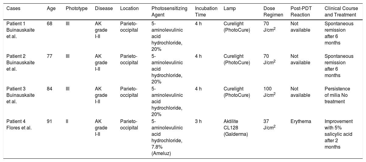
The appearance of milia following PDT is a rare adverse effect that we had not previously observed in our extensive clinical experience. This phenomenon is known to reflect the severity of phototoxic damage and disruption of the dermal-epidermal junction.4 Milia have been reported in the literature in 3 patients 3 months after PDT (5-aminolevulinic acid hydrochloride). In 2 of the cases, spontaneous resolution was observed after 6 months5 (Table 1).

Table 1.

Cases of Milia Following Photodynamic Therapy Published in the Literature.

Cases  Age  Phototype  Disease  Location  Photosensitizing Agent  Incubation Time  Lamp  Dose Regimen  Post-PDT Reaction  Clinical Course and Treatment 
Patient 1
Buinauskaite et al. 
68  III  AK grade I-II  Parieto-occipital  5-aminolevulinic acid hydrochloride, 20%  4 h  Curelight (PhotoCure)  70 J/cm2  Not available  Spontaneous remission after 6 months 
Patient 2
Buinauskaite et al. 
77  III  AK grade I-II  Parieto-occipital  5-aminolevulinic acid hydrochloride, 20%  4 h  Curelight (PhotoCure)  70 J/cm2  Not available  Spontaneous remission after 6 months 
Patient 3
Buinauskaite et al. 
84  III  AK grade I-II  Parieto-occipital  5-aminolevulinic acid hydrochloride, 20%  4 h  Curelight (PhotoCure)  100 J/cm2  Not available  Persistence of milia No treatment 
Patient 4
Flores et al. 
91  II  AK grade I-II  Parieto-occipital  5-aminolevulinic acid hydrochloride, 7.8%
(Ameluz) 
3 h  Aktilite CL128 (Galderma)  37 J/cm2  Erythema  Improvement with 5% salicylic acid after 2 months 

Abbreviations: AK, actinic keratosis; PDT, photodynamic therapy.

In our case, in contrast to the 3 cases mentioned above, onset of milia occurred earlier and the condition resolved more quickly following keratolytic treatment. This difference may be linked to the severity of the inflammatory reaction our patient presented after treatment. As in the cases mentioned above, the milia appeared following the application of topical 5-aminolevulinic acid hydrochloride in actinic keratosis. No similar reactions have been reported to date in cases in which the photosensitizing agent was methyl aminolevulinate. This phenomenon may be explained by the greater affinity of 5-aminolevulinic acid hydrochloride for cancerous tissue,6 which leads to greater collateral damage of healthy tissue.

Paradoxically, PDT is one of the most commonly used treatments for milia en plaque.7

We present a new case of milia following treatment of actinic keratosis using PDT. The patient had no past personal or family history of milia. Given the benign and potentially self-limiting nature of the condition, it is important for dermatologists to be able to identify milia and avoid mistaking it for treatment failure. In our case, although we explained that the condition was benign, the patient stated that he was unhappy with the cosmetic result and we therefore started mild keratolytic treatment with very good results.

Conflicts of Interest

The authors declare that they have no conflicts of interest.

References
[1]
D.R. Berk, S.J. Bayliss.
Milia: a review and classification.
J Am Acad Dermatol., 59 (2008), pp. 1050-1063
[2]
A.M. Pisauri, A. Alvarez-Gracia, C. Ferrandiz-Foraster, J. Bassas-Vila.
Milia en placa en la cara posterior de ambos pabellones auriculares secundaria a radioterapia.
Actas Dermosifiliogr., 107 (2016), pp. 156-158
[3]
L.M. Miller, J.T. Schwartz, S. Cho.
Milia: a unique reaction to tattoos.
Cutis., 87 (2011), pp. 195-196
[4]
S.H. Ibbotson.
Adverse effects of topical photodynamic therapy.
Photodermatol Photoimmunol Photomed., 27 (2011), pp. 116-130
[5]
E. Buinauskaite, R. Maciulaitis, J. Buinauskiene, S. Valiukeviciene.
Topical photodynamic therapy of actinic keratoses with 5-aminolevulinic acid: randomized controlled trial with six months follow-up.
J Dermatolog Treat., 25 (2014), pp. 519-522
[6]
C. Sandberg, J. Paoli, M. Gillstedt, C.B. Halldin, O. Larkö, A.M. Wennberg, et al.
Fluorescence diagnostics of basal cell carcinomas comparing methyl-aminolevulinate and aminolaevulinic acid and correlation with visual clinical tumour size.
Acta Derm Venereol., 91 (2011), pp. 398-403
[7]
M.P. Stefanidou, J.G. Panayotides, A.D. Tosca.
Milia en plaque: a case report and review of the literature.
Dermatol Surg., 28 (2002), pp. 291-295

Please cite this article as: Flores-Climente V, Dalmau J, Puig L. Milia, una reacción infrecuente tras terapia fotodinámica. Actas Dermosifiliogr. 2019;110:71–74.

Copyright © 2018. Elsevier España, S.L.U. and AEDV
Idiomas
Actas Dermo-Sifiliográficas
Opciones de artículo
Herramientas
es en

¿Es usted profesional sanitario apto para prescribir o dispensar medicamentos?

Are you a health professional able to prescribe or dispense drugs?