


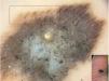
An 80-year-old woman presented with a pigmented lesion on her left plantar arch of uncertain duration, which had been brought to her attention by her podiatrist. Examination revealed a flat pigmented plaque measuring 2cm, with irregular borders, dark brown coloration at the periphery, and a gray-blue center. Dermoscopy showed a multicomponent pattern consisting of a parallel ridge pattern, a blue-white veil, and atypical globules that were notably arranged along the dermatoglyphic ridges at the periphery of the lesion. Histopathology revealed a non-ulcerated melanoma with a Breslow thickness of 1.6mm.
The dermoscopic pattern classically associated with acral melanoma is the parallel ridge pattern, but other findings may also be present, such as diffuse pigmentation, multicomponent patterns, fibrillar patterns, or irregular lattice patterns. Histopathologically, the globules observed on dermoscopy correspond to nests of melanocytes located at the dermoepidermal junction or being eliminated through the stratum corneum. Typically, atypical globules with irregular distribution are found in melanomas; however, a distinctive variant has been described in which globules are specifically distributed along the ridges, referred to as the “irregular globular pattern predominantly located on the ridges.” The main differential diagnosis is congenital nevi, in which globules are regularly arranged and may be accompanied by a parallel sulcus pattern, forming the so-called “peas-in-a-pod pattern.” (Fig. 1).


