


Tattooing has been practiced for more than 8000 years. During the last 30 years, the practice has spread widely in developed countries.1
Complications of tattooing include transmission of infectious diseases (mainly mycobacterial diseases, which are often associated with the color gray, through use of nonsterile water as a diluent), underlying skin diseases resulting from an isomorphic process, and even tumors at the site of the tattoo. Furthermore, injecting a foreign substance into the skin can induce a toxic or immune response.2
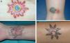
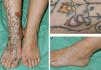
Table 1 presents the characteristics of the study patients. The first case involved a woman who experienced intense itching and raised red areas after receiving a multicolored tattoo (Fig. 1). The following patient, who had granuloma annulare as a child, experienced itching in the red areas of a multicolored tattoo (Fig. 2A), in which some parts were raised and others eroded, with production of material (Fig. 2B). Brownish maculopapular lesions appeared 1 month later, taking on an irregular circinate pattern on the dorsum of the foot and distal end of the lower left leg (Fig. 2C). Histopathology of the lesions revealed granuloma annulare. The third patient had received a green and red tattoo, which progressed with subcutaneous nodules that became ulcerated on the red areas (Fig. 2B). The fourth patient complained of itching and inflammation on the red areas of a tattoo placed on her right wrist 15 days earlier. The inflammation progressed to forma a large ulcer (Fig. 1C). Five months after the onset of symptoms on the wrist, the patient also complained of inflammation and discomfort on a tattoo that had been placed 5 years previously (Fig. 1D).
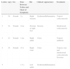
Characteristics of 5 Lesions in 4 Patients.
| Lesion | Agey | Sex | Time Between Tattoo and Onset of Symptoms | Site | Clinical Appearance | Treatment |
|---|---|---|---|---|---|---|
| 1 | 28 | Female | 2 y | Right forearm | Erythema/Inflammation | Topical corticosteroids |
| 2 | 24 | Female | 4 mo | Dorsum of right foot | Erosion | Intralesional corticosteroid |
| 3 | 23 | Female | 1 mo | Right ankle | Ulcer | Topical antibiotic |
| 4 | 38 | Female | 15 d | Right wrist | Ulcer | Topical, oral, and intralesional corticosteroids. Excision |
| 5 | >5 y | Left ankle | Erythema/Inflammation |
A, Lesion No. 1: The image shows inflammation of the red areas in a multicolored tattoo. B, Lesion No. 3: The image shows ulceration and inflammation on the red area of a red and green tattoo. C, Lesion No. 4: The image shows a large ulcer in the red areas of a green, red, and black tattoo. D, Lesion No. 5: The image shows inflammation of the red area of a tattoo that first appeared 5 months after the appearance of lesion No. 4 in the same patient.

Histopathology of the 5 lesions revealed an inflammatory reaction with multiple multinucleated giant cells and pigment compatible with foreign body granuloma (Fig. 3). The results of microbiology studies based on staining and culture (including mycobacteria) were negative for all 5 lesions. In the second case, systemic sarcoidosis was ruled out by chest x-ray and determination of angiotensin-converting enzyme in blood.
![Histopathology study of lesions resulting from a granulomatous reaction to a tattoo. Granulomas can be observed in the superficial dermis (A, hematoxylin-eosin [H-E] ×4) and deep dermis (B, H-E ×4). Greater magnification reveals foreign body granulomas around the pigment (C, H-E ×10) and phagocytic cells filled with red pigment (D, H-E ×40).](https://static.elsevier.es/multimedia/15782190/0000010600000007/v1_201508271313/S1578219015001808/v1_201508271313/en/main.assets/gr3.jpeg?xkr=ue/ImdikoIMrsJoerZ+w91sAmkCw32Jed9sZf6jzEuDbFpW7G0NfARZs8afh+9K8v8RN+oFy2ZmalFHXVYo6AkGUq0SAScLCI7yrq7jOeaehRlF8Sr1y6gGEdt3Gamh5o0Vzr4lms39bTsflIcxmiS4QC+LuaC1FcT+qjCCzLjrNeH/vPnn1l2oKYX4VE17oKiTvaO8EkLv54Tr0xPvAemZwjc4OVc0E0+EYyN48HKOD2rtAoR+4D5tMGniAu6XWR8UuY/raC6kBPshY9GnDALZJoLdhWW9MgHvjKIqegEQ=)
Histopathology study of lesions resulting from a granulomatous reaction to a tattoo. Granulomas can be observed in the superficial dermis (A, hematoxylin-eosin [H-E] ×4) and deep dermis (B, H-E ×4). Greater magnification reveals foreign body granulomas around the pigment (C, H-E ×10) and phagocytic cells filled with red pigment (D, H-E ×40).
Adverse cutaneous hypersensitivity reactions to tattoos are not uncommon and are attributed to the materials injected. Red is the color most commonly associated with adverse reactions. Red tattoo pigments were traditionally made from mercury derivatives, then from cadmium derivatives, which are currently prohibited because of toxicity.3,4 Today, tattoos contain synthetic organic pigments, which carry a lower risk of adverse reactions. The problem is that there are no strict regulations on pigment content, and most professional tattoo artists do not know the composition of the pigments they use.1 People receiving a tattoo are for the most part unaware of the substances being injected and of their possible adverse effects. In 2008, the Spanish Agency for Medications and Medical Devices drafted a list of authorized registered products that should not exceed the maximum limits for harmful substances, such as mercury, lead, and arsenic.5 In any case, the exact composition of each of the products used is unknown.
The histopathologic patterns produced by adverse reaction to a tattoo are diverse, the most frequent being a lichenoid pattern. Other types of reactions to tattoos include eczematous, pseudolymphomatous, and sclerodermiform reactions, as well as perforating collagenosis, and, as in the cases we report, granulomatous reactions.6
Although very rare, sarcoid granulomas can be found within a tattoo. This finding can be the only clinical manifestation of systemic sarcoidosis.7
We observed 1 case of granuloma annulare that occurred simultaneously with and at a distance from a granulomatous reaction to red tattoo pigment. The literature contains several cases of granuloma annulare limited to the red areas of the tattoo,8 but not distant lesions. The etiologic and pathogenic mechanisms of granuloma annulare remain unknown, although it is postulated that altered collagen and elastic fibers could trigger the immune response.9
Cutaneous reactions to tattoo pigment are treated mainly with topical corticosteroids. Reactions to red pigment have been treated with Q-switched Nd:YAG laser, which carries a risk of severe allergic reactions resulting from lysis of phagocytes and release of pigment into the extracellular space.10
The increased frequency of tattooing in our setting means that dermatologists should be aware of potential side effects and able to diagnose them correctly. We believe that people who wish to have a tattoo should be informed of the risk of severe reactions, especially when red pigment is used.
Please cite this article as: Martín-Callizo C, Marcoval J, Penín RM. Reacciones granulomatosas a los tatuajes rojos: presentación de 5 lesiones. Actas Dermosifiliogr. 2015;106:588–590.


