


One of the main strategies in the prevention of photodermatoses is the use of topical sunscreens, which sometimes must be applied together with topical drugs specific for the dermatosis with the possibility that the efficacy of the sunscreen may be altered.
ObjectivesThe aim of this study was to analyse whether the efficacy of the sunscreen could be affected when applied together with topical drugs routinely used in patients with photodermatoses and whether there is any variation when they are applied before or after the drug.
MethodsNinety-three volunteer patients with photodermatoses participated. Very high SPF sunscreens were used with corticosteroids, antibiotics and topical antifungals as active ingredients. Paravertebral areas were delimited in each individual and the sunscreen was applied alone and in association with a drug in different sequential order. Ultraviolet reflectance photography and image analysis were used to compare the level of UV absorption by the sunscreen/drug combinations in the different areas.
ResultsUV reflectance analysis showed no difference in the efficacy of the sunscreen applied before or after the various drugs used. In the antifungal group, a significant increase in the effect of the sunscreen was observed when the antifungal was applied first.
ConclusionsThe efficacy of sunscreens was not altered by combined use with corticosteroids, antibiotics or topical antifungals. These results are of great relevance for patients with photodermatosis who often have to combine these active ingredients with photoprotection.

Photodermatoses are skin diseases induced or exacerbated by electromagnetic radiation (including UV radiation, visible light, and infrared radiation) emitted by the sun or artificial sources.1 Patients with photodermatoses develop cutaneous lesions after minimal doses of light irradiance, and in severe cases, artificial lighting may be a contributing factor as well. This explains why the adoption of preventive photoprotection measures is an essential pillar in their therapeutic management.1–3
One of the most effective measures to prevent the adverse effects of sun exposure is the use of topical sunscreens. These products are characterized by the presence of compounds capable of absorbing or reflecting UV radiation, thereby preventing its penetration into the skin.4,5 Sunscreens are highly stable and provide broad protection against UVA and UVB rays. Their use is complemented by other measures such as wearing sunglasses, dark clothing, and hats to avoid direct exposure to radiation.4,5
Most patients with photodermatoses, in addition to adopting comprehensive photoprotection measures, require topical therapies to protect their underlying condition from UV overexposure.1–3,6,7 Among these medical therapies, topical corticosteroids are most widely used ones as first-line therapy in conditions such as polymorphic light eruption and other inflammatory dermatoses, including solar urticaria, cutaneous lupus erythematosus, chronic actinic dermatitis, and even lesions produced as an immune response to exogenous substances that may cause photoallergic reactions. On the other hand, topical antibiotics such as fusidic acid, mupirocin, and gentamicin are useful in superinfected lesions, while others such as erythromycin or metronidazole are used in patients with rosacea, as well as in those affected by photo-aggravated eczema.8
It is common for patients with photodermatoses to apply both topical treatments for their condition and sunscreens simultaneously or within a short time interval; however, it has not yet been established whether this combination could affect sunscreen efficacy. To address this question, UV photography has been used as an ideal non-invasive tool. Based on the physicochemical properties of sunscreens – specifically their absorption of UV radiation – it is possible to quantify the degree of absence of reflection of incident light (in this case, UV). Consequently, skin images obtained in the presence of sunscreen appear dark or black. This technique has been used in several research studies as a means of raising awareness regarding sunscreen use by visualizing skin with and without photoprotection,9–11 to indicate correct sunscreen application in terms of quantity12 and body area,13 and to determine the time required for sunscreen to become effective on the skin,14 either alone or combined with moisturizing formulations.15
The aim of the present study was to determine, using UV photography, potential alterations in the efficacy of topical sunscreens when applied to the same skin area together with topical medications.
Materials and methodsStudy design and study populationWe conducted an observational, cross-sectional, and descriptive study to evaluate the UV radiation absorption potential of topical sunscreen formulations before and after the application of topical drugs widely used by patients with photodermatoses. All patients participating in the study were diagnosed with photodermatoses and were recruited from three hospitals from the province of Málaga (Spain). The study was conducted at the Dermatological Photobiology Laboratory of the Medical and Health Research Center of Universidad de Málaga (Málaga, Spain). All volunteers gave their prior written informed consent to participate after being properly informed in full compliance with the ethical principles outlined the Declaration of Helsinki. The study was approved by the Ethics Committee of the Andalusian Regional Ministry of Health.
All patients underwent a photobiological clinical history assessment to document the clinical course of their photodermatosis and determine the appropriate photodiagnostic protocol based on their prior clinical suspicion (erythema phototesting, abnormal UVA response, photoprovocation, photopatch testing). Twenty-seven of all patients were diagnosed with solar urticaria (29.03%), 21 with photosensitivity to exogenous agents (22.58%), 14 with idiopathic photosensitivity (15.05%), 11 with polymorphic light eruption (11.83%), 7 with contact dermatitis (7.53%), and 5 with tumid lupus erythematosus (5.38%); the remaining 8 patients exhibited photodermatoses of unknown etiology (8.60%).
Volunteers were categorized into three main groups according to the type of topical therapy used. In group #1 (37 patients), corticosteroids were studied (methylprednisolone aceponate 0.21%, beclomethasone dipropionate 0.025%, prednicarbate 0.25%, hydrocortisone aceponate 0.127%, and betamethasone valerate 0.05%). In group #2 (31 individuals), antibiotics were evaluated (mupirocin 2% and fusidic acid 2%), and in group #3 (25 patients), antifungals were assessed (ketoconazole 2%, clotrimazole 1%, and bifonazole 1%). As this experimental design compared sunscreen efficacy against itself, each subject served as their own control.
Product application protocol and image acquisitionThe procedure was performed on the dorsal region of each patient, selecting a paravertebral area in which both sunscreens and topical drugs provided by the patient were tested. In cases where the patient did not provide either drug or sunscreen, commercially available very high SPF (50+) sunscreens were tested, along with the P8 standard used as a reference for very high SPF category in the ISO 24444:2019 sunscreen protection factor calculation assay,16 as well as different commercially available drugs from each one of the three categories.
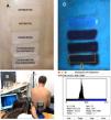
Five 5cm2×2cm2 rectangular areas were delineated (Fig. 1) using a plastic template fixed to the skin for product testing: (1) untreated skin, (2) sunscreen, (3) medication, (4) sunscreen (10min) followed by medication, and (5) medication (10min) followed by sunscreen. Sunscreen application followed the recommendations of Cosmetics Europe and the ISO 24444:2019 standard (2mg/cm2), as did the application of each topical drug. Ten minutes after the application of the different product combinations in each area, the delimiting template was removed and UV photography was performed.

(A) Left paravertebral region divided into five 5cm2×2cm2 areas with the respective treatments applied. (B) UV photography device used to capture images of each volunteer's back. (C) UV photograph showing treated areas and the rectangular region selected for pixel analysis using Fiji–ImageJ software. (D) Histogram showing pixel color distribution of the selected image area, allowing comparison of color levels (mean pixel value) for each treatment.
UV image acquisition was conducted following a previously described protocol,13,14 using a Canon EOS 500D digital reflex camera (Canon Co., Tokyo, Japan) equipped with 2 halogen flashes and 1 Schott UG11 interference filter (Schott AG, Jena, Germany). A Schott BG38 filter was placed in front of the lens to eliminate residual visible light. Camera control and image acquisition were performed using Canon EOS Utility 2 software (shutter speed=1/20, aperture=f/5.0, ISO=1600).
Image analysisUV photographs were analyzed using Fiji–ImageJ software, an open-source program (GNU General Public License) (Fig. 1).14 A color histogram was generated based on the black–blue pixel tone intensity provided by the image, using a color scale ranging from 0 to 255 (0=black, 255=lightest tone on the blue–black scale). The obtained color levels were evaluated in terms of percentage changes in color reduction relative to untreated skin.
Statistical analysisFor statistical analysis, mean values of the percentage changes and their corresponding 95% confidence intervals were calculated. The results of the analyzed areas were compared using one-way analysis of variance (ANOVA) for each drug type, followed by Bonferroni post hoc testing for comparisons across treatment types. Differences were considered statistically significant when p<0.05. Statistical analysis was performed using IBM SPSS version 20.
ResultsThe final sample consisted of 93 patients aged 18–70 years (65% women/35% men), with 35% phototype II and 65% phototype III. All sunscreens used – both those routinely applied by patients attending the photodiagnosis clinic and those supplied by the research group for testing – were obtained from different commercial manufacturers and international standards. All products had a sun protection factor (SPF) of 50 or 50+. No specific classification of sunscreens was conducted for the study, since all tested products showed a similar behavior on the skin, with a significant and comparable decrease in color level (reductions of 39–45% on the blue/black scale), which is consistent with their equivalent SPF levels. Therefore, the analysis of results was structured according to the groups of concomitant topical drugs (corticosteroids, antibiotics, and antifungals).
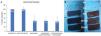
CorticosteroidsWhen analyzing the mean results of all corticosteroids together (Fig. 2A), the area where sunscreen was applied showed a mean reduction in color level from 100% to 47.6% (95% CI, 38.2–57%). When the topical corticosteroid was applied before the sunscreen, color reduction reached 44.1% (95% CI, 34.8–53.5%), and when applied after the sunscreen, 47.4% (95% CI, 38.8–56%). No statistically significant differences were observed vs sunscreen alone (p>0.05).

Distribution of mean percentage color-level values for different treatment areas relative to untreated skin. (A) Mean color-change values grouping all corticosteroids used. (B and C) Examples of color distribution in two different volunteers treated with corticosteroid and SPF 50+ sunscreen.
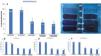
Similar results were observed in the antibiotic group. Application of mupirocin alone showed a slight reduction in the white-to-black color level from 100% to 90.4% (95% CI, 85.9–94.9%). Although these differences were not statistically significant compared with untreated skin (p>0.05), mupirocin and fusidic acid results were grouped for analysis (Fig. 3D–F). Application of sunscreen alone reduced the color level from 100% to 42.1% (95% CI, 33.1–42.8%). This effect was not altered when the antibiotic was applied before the sunscreen (drop down to 36.5%; 95% CI, 30.9–42.1%) or after sunscreen application (drop down to 37.2%; 95% CI, 33.1–42.8%) (Fig. 3A).
Antifungals
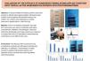
In the antifungal group, bifonazole absorbance (Fig. 3D) showed a significant color reduction to 85% (95% CI, 89.2–89.8%) vs untreated skin (example image in Fig. 3C). In contrast, ketoconazole (example in Fig. 3B) and clotrimazole did not show significant changes. When antifungals were grouped, application before sunscreen reduced color level to 39.3% (95% CI, 33.7–44.8%) relative to untreated skin, while sunscreen alone produced a reduction to 49.38% (95% CI, 39.8–53.6%) (Fig. 4A). When the antifungal was applied after sunscreen, the reduction in color percentage was not significantly different from sunscreen alone (p>0.05).

Distribution of mean percentage color-level values for different treatment areas relative to untreated skin. (A) Mean color-change values grouping all antifungals used. (B and C) Examples of color distribution in two volunteers treated with antifungal and SPF 50+ sunscreen. (D–F) Mean percentage color-level values for all volunteers treated with ketoconazole 1%, clotrimazole 1%, and bifonazole 1%. *Significant differences (p<0.05) between sunscreen alone and antifungal applied before sunscreen. **Significant differences between bifonazole alone and untreated skin.
This study demonstrates that the UV-blocking and/or reflective capacity of sunscreens is not altered when applied either before or after topical drugs belonging to the corticosteroid, antibiotic, or antifungal classes. Moreover, when antifungals were applied prior to sunscreen, a further reduction in color level was observed, indicating increased UV absorption. Therefore, topical photoprotection – when applied according to international recommendations on dry skin at a concentration of 2mg/cm2 and reapplied every 2h – can safely be used concomitantly with topical medications without compromising sunscreen efficacy.16 In addition, as previously demonstrated, it is not necessary to wait 30min after sunscreen application before sun exposure.15
One of the limitations of this study is that, although widely used drugs for frequent dermatoses were tested, it does not include the full range of topical formulations available on the market. The vehicle of the photoprotective products was taken into consideration when designing the study, as a significant variety of formulations with different lipid contents and textures (ranging from sprays to creams) were used. In the case of topical drugs, all products employed were typical high-oil-in-water (o/w) cream formulations, as is customary for these agents. Regardless of the galenic formulation of the photoprotectors, because all were classified as SPF 50 or 50+, their UV absorption behavior for image acquisition did not differ significantly. Moreover, this study assessed the efficacy of the photoprotective product. However, to objectively determine whether the use of photoprotection could alter drug efficacy, a different type of study would be required, as image analysis alone is insufficient. Assuming that concomitant use does not affect the efficacy profile of either topical formulation, the recommended order of application should be as follows: first, apply the topical drug to clean, dry skin in the appropriate amount and coverage to ensure adequate absorption, and after several minutes (>10min), apply the photoprotective product. Very similar results emphasizing the use of one topical substance over another have been published previously, showing that the application of moisturizing formulations for cosmetic purposes did not affect the efficacy of photoprotectors.15
The use of UV photography in the field of photoprotection has been employed for a considerable time as a method to observe skin damage, as well as a tool to raise awareness on the use of different photoprotective strategies for the prevention of photoaging and the long-term development of skin cancer.9,10,17–19 Photographic image analysis has enabled greater precision in the study of the behavior of topical photoprotectors on the skin, allowing visualization of correct application and persistence.11–13,20 In the present study, observation by patients attending the photodiagnosis clinic of the UV absorption effect of photoprotectors promoted awareness of sun protection, particularly for the prevention of their own cutaneous lesions.
The final conclusion of this study was that the efficacy profile of photoprotectors was not altered by combined use with topical corticosteroids, antibiotics, or antifungal agents, regardless of whether these were applied before or after photoprotection. These findings are highly relevant for patients with photodermatoses, who frequently need to combine these active agents with photoprotective measures.
ConclusionsThe efficacy profile of sunscreens was not altered by combined use with topical corticosteroids, antibiotics, or antifungals. These findings are highly relevant for patients with photodermatoses, who frequently need to combine these active ingredients with photoprotection.
FundingThis research was funded by the Spanish Ministry of Science and Innovation, State Program for Knowledge Generation and Scientific and Technological Strengthening (Grant No. PID2020-117224RB-100). This work forms part of the research activities of the Institute of Biomedicine of Málaga (IBIMA) and the Andalusian Regional Government research group CTS-162.
Conflict of interestThe authors declare no conflict of interest.


