


Ectodermal dysplasias are rare major inherited disorder characterized by changes in structures of ectodermal origin. The syndrome is characterized by ectrodactyly, hypoplasia of the mammary glands and nipples, cleft palate, tear duct stenosis, hearing loss, urogenital abnormalities, nasal dysplasia, hypohidrosis, hypodontia, gonadaldysplasia, and absence of skin or hair abnormalities.1,2 Possible causes of congenital breast asymmetry; it may be due to a developmental or hormonal problem, a syndrome, to familial or a genetic disorder. In this manuscript, we describe a family with different clinical findings over four generations.
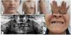
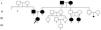
A 14-year-old girl presented complaining about the underdevelopment of the right breast (Fig. 1B, C). The clinical examination showed a normal nipple but with no normal left breast tissue (E-5) and no areola in the width of the nipple. The right pectoralis major muscle was normal. She also had facial palsy (Fig. 1A), extensively depigmented skin areas, hyperpigmentation, irregular and missing teeth (Fig. 1E), hearing loss, pubic hair P4-5, and slightly elongated axillary hair (A-2). External genitalia, uterus, and ovaries were normal, but there was no cliteromegaly. She also had small hands (Fig. 1D), closed epiphysis, atrophic left kidney, and endocrinological problems. Hormones such as FSH, LH, E2, prolactin, progesterone, testosterone, TSH, FT4, mental and motor development were normal. Father and mother were diagnosed with obsessive compulsive disorder and mild mental retardation. Physical dental problems (Fig. 1F) and speech disorders were observed in the parents. It was also reported that the sweat glands of the mother lost their function during childhood, that she had goiter, allergic problems and learning difficulties, and that the father's hair did not grow and was abnormal. Consanguinity (I-3,4), learning, mild mental retardation, nervous disorders and biliary disorders were recorded in the first generation grandparents. However, congenital hearing loss was also found in two other members of the third generation (Cousins, III-5,6) (Fig. 2).

14-Year-old patient with a hypoplastic breast (the small right breast) (B, C), the small left nipple (B, C), facial paralysis (A), small hands (D), missing teeth (E), and mother's teeth (F) (Fig. 2).

It was determined that 9 permanent teeth of the patient were still in the growth process and 4 primary teeth were still in place. It was recorded that the permanent teeth of the patient were left upper anterior central incisors, right upper and left upper canines, right upper and left upper 1st molars, lower right and lower left 1st molars and upper left 2nd molars. The canines had dysmorphological features such as the morphology of the nail-shaped incisors tapering toward the edges, and the other permanent teeth had normal morphological features. Retained teeth and teeth losses in patient were not detected. One deciduous molar for each half jaw and bone loss in the lower anterior part due to edentulism were observed (Fig. 1E).
The cytogenetic results of our patient showed a normal chromosomal complement. However, structural and numerical chromosome changes were detected in 22.5% of the mother cells.
The clinical and phenotypic findings of the patient's parents were also consistent with EDs. In addition, it was reported that the mother and father of the mother (Grandparents, I-3,4) were consanguineous and both had similar clinical findings. As a matter of fact, it was recorded that both daughters of the uncle of the proband were congenitally deafness (Cousins, III-5,6). The familial history shows us that the clinical findings in the proband are hereditary. Cases of asymmetrical breasts have been reported in some families before. In one of these families, bilateral breast absence was reported in 1 boy, 3 out of 4 girls and 3 grandchildren over three generations.3 On the other hand, in a family where the mother and father were not affected, male and female cousins did not have bilateral breasts in another family, 7 individuals in 4 generations were reported to have no breasts or hypoplastic breasts.4,5 Our findings suggest that the presence of asymmetric breasts is a serious and rare pathology of ED.
Since the otic capsule is of ectodermal origin, it is natural to see sensorineural hearing loss in ED. In the current family, three members of the third generation (probant, cousins, III-1,5,6) had congenital hearing loss. It is noteworthy that all three of these cousins were female. The hearing loss in our patients was consistent with the previous case series.6 Intelligence, motor and cognitive development were found to be affected in some other members of the family. Some studies have reported abnormal motor/mental development in 15–25% of patients affected by HED.7 Various renal problems have been identified in individuals affected by ED. We have reported atrophic kidney structure in the patient. The mother of the patient had many structural and numerical irregularities in different chromosomes. It should be kept in mind that structural damages in the 2nd chromosome (q11-ter, q14, q21, q23 and p23 regions) and 19, 21 and other chromosome losses detected in the mother may be associated with ecdodermal tissue development. A gene responsible for autosomal HED has recently been mapped to chromosome 2q11-q13 in human and mouse.8 This gene has also been shown to be expressed in the epithelium and in adjacent or partially overlapping layers of developing human skin.9 All this information shows that chromosome 2 plays a role in the etiology of ED.
Conflict of interestThe authors declare they have no conflict of interest.
We gratefully thank the Department of Pediatrics for referring the family to our department. I would like to express my gratitude to the aunt, who took close care of the index patient and her mother, contributed to the acquisition of information, and filled out the consent form in line with their approval.


