


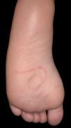
A 3-year-old boy presented with a 2-week history of a skin lesion on the plantar surface of the left foot. The lesion was mildly painful when walking but did not cause pruritus. There was no history of recent travel or associated systemic symptoms. Interestingly, his parents reported that the lesion appeared to progress gradually and that he often walked barefoot. Physical examination revealed a curvilinear plaque composed of a central gray-black line with slight surrounding erythema on the left plantar surface (Fig. 1). Dermoscopy showed a well-defined central black, filiform structure extending along the entire lesion. A hair was extracted from the lesion by gentle curettage at its distal end with the aid of forceps, confirming the diagnosis of cutaneous pili migrans and resulting in complete resolution.

With approximately 50 published cases, cutaneous pili migrans is characterized by skin lesions consisting of a linear hair shaft that is mobile and may be accompanied by erythema. Reported sites include the ankles, soles, toes, breasts, cheeks, neck, and abdomen, with the soles being most common in children and the abdomen in adults. Although dermoscopy is usually helpful in visualizing the causative hair shaft within the epidermis, biopsy may be necessary in some cases because the hair lies deeper in the dermis, and the lesion may present only as an elevated erythematous area. The absence of pruritus or travel history, the visualization of a hair shaft, and the unidirectional migration of the lesion are key features that differentiate pili migrans from cutaneous larva migrans and help avoid unnecessary treatments. Another important differential diagnosis is barber's interdigital pilonidal sinus, an occupational condition characterized by immobile inflammatory lesions in the interdigital spaces that may progress into fistulas due to penetration of foreign hair and a foreign-body reaction. Treatment is usually simple, consisting of removal of the hair with forceps after a small incision or curettage.


