


The Mpox outbreak (previously known as monkeypox) that began in May 2022 has epidemiological and clinical features that differ from those described previously. Sexual contact is the main route of transmission, predominantly among MSM (men who have sex with men), and therefore the anogenital and perioral regions are frequently affected.1 Below, we describe two illustrative cases of phimosis secondary to Mpox – an uncommon form of presentation.
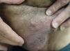
A 50-year-old man presented to the emergency department with a 1-week history of edema due to an ulcerated penile lesion. The patient reported recent high-risk sexual contact (unprotected oral sex with another man) and had experienced 15 days before presentation a non-specific systemic illness with malaise, low-grade fever, myalgias, upper airway congestion, and anorexia. The penile lesion appeared the day after these systemic symptoms resolved. Physical examination highlighted marked edema of the glans and foreskin that prevented retraction, along with a single ulcer on the preputial ring with a fibrinous base, indurated to palpation and non-tender, without palpable lymphadenopathy (Fig. 1). The main working diagnosis was primary syphilis. A full syphilis serology and STI screening from urethral exudate were requested. Treponemal tests tested positive, but RPR tested negative, and the patient acknowledged having previously been treated with penicillin. Given the negative results and persistent suspicion, PCR testing of the ulcer exudate was requested for Herpes simplex virus, Treponema pallidum (still considered the main suspicion), and Mpox – the latter added because significant edema has been described as a complication of this infection. The PCR result came out positive for Mpox, and the patient was advised to isolate. Oral prednisone was prescribed to reduce the edema responsible for the phimosis.

A 68-year-old man, HIV-positive, also presented to the emergency department with similar swelling of the glans and foreskin due to a lesion on the free margin of the foreskin, with an inability to retract the skin over the glans. He had engaged in unprotected MSM sexual activity a few days before the onset of symptoms (Fig. 2). In this case, diagnosis was easier due to the presence of multiple typical lesions elsewhere in the genital area (Fig. 3); PCR for Mpox was also positive. Screening for other STIs revealed latent syphilis.


In the current Mpox outbreak, the most frequent location of skin lesions is the anogenital region, and secondary genital edema has been described as a relatively common complication (8–16% of reported cases2), potentially resulting in phimosis3,4 or paraphimosis,5 with phimosis more frequently observed in patients coinfected with HIV.6 Moreover, in up to 11% of cases,7 Mpox presents as a single lesion, which can lead to diagnostic confusion, especially with syphilitic chancres. In such situations, prominent genital edema can be a helpful distinguishing feature. It is also important to highlight the high rate of coinfection with other sexually transmitted infections – seen in more than half of cases8 – which makes comprehensive screening for additional STIs essential.
Conflict of interestThe authors declare no conflict of interest.


