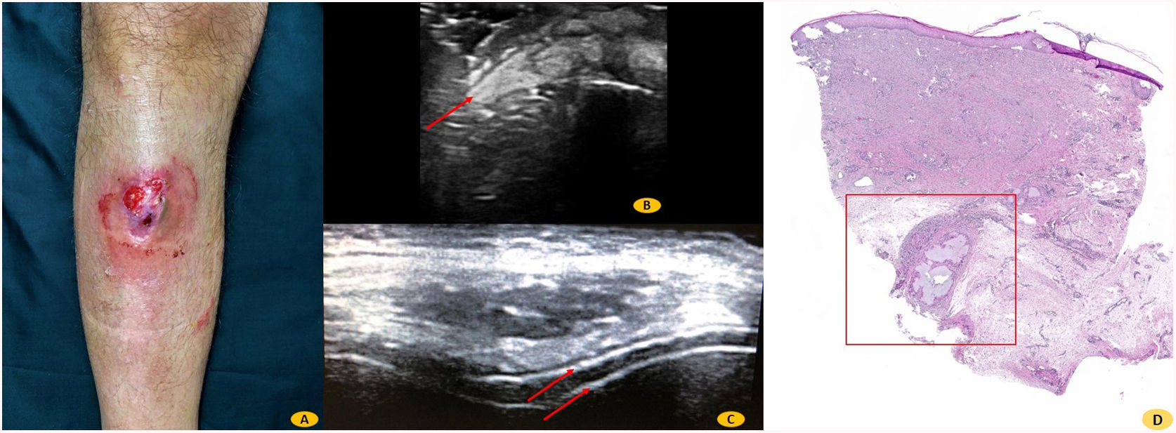


A 53-year-old man with a history of gout was evaluated in the dermatology department for the presence of an ulcerated, densely friable nodule with a deposit of hard white material on the surface, without purulent discharge (Fig. 1A). The nodule had appeared 6 months earlier. Soft tissue ultrasound revealed a heterogeneous mass with a lobulated border and hyperechoic trabecular structures (Fig. 1B). Ultrasound examination of the right knee revealed minor effusion and the presence of the double contour sign, which is considered highly suggestive of urate deposition in the hyaline cartilage (Fig. 1C). Histology showed amorphous basophilic material surrounded by a foreign-body-like giant-cell reaction with focal lymphohistiocytic infiltrate (Fig. 1D). Uric acid levels were 9.5mg/dL (3.4–7.0mg/dL). All other laboratory parameters were within the normal range. In terms of the pathophysiology of gouty panniculitis, venous stasis and pre-existing tissue damage are factors that, together with repeated microtrauma, make patients more predisposed to urate crystal deposition on subcutaneous tissue. Serum uric acid levels do not appear to be directly related to the development of panniculitis, as was previously suggested.
Impact factor
The Impact Factor measures the average number of citations received in a particular year by papers published in the journal during the two preceding years.
© Clarivate Analytics, Journal Citation Reports 2022
Impact factor 2023
3.8
Citescore 2023
1.9
SJR
SRJ is a prestige metric based on the idea that not all citations are the same. SJR uses a similar algorithm as the Google page rank; it provides a quantitative and qualitative measure of the journal's impact.
See moreSJR 2023
0.30
SNIP
SNIP measures contextual citation impact by wighting citations based on the total number of citations in a subject field.
See moreSNIP 2023
0.68
View more metrics