


We present the case of a fifteen-year old boy who presented with a history of hypertrophic nail dystrophy and subungual debris of all 20 nails dating back to childhood. He also suffered plantar pain with occasional painful bullae and fissures at friction sites, as well as multiple palmoplantar infections which had been treated with oral antibiotics and topical antifungals. His mother presented the same clinical condition but to a lesser degree, and no other family members were affected. They did not report erupted teeth at birth, hearing loss or hair disorders.
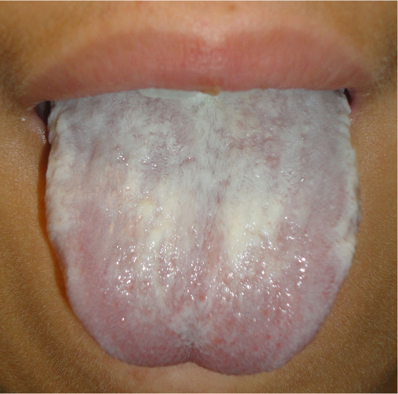
Physical examinationOn examination, all his fingernails and toenails were dystrophic, discolored, and thickened, with massive subungual hyperkeratosis (Fig. 1a). Keratotic plaques and bullae following pressure distribution were found on the palms and soles (Fig. 1b), and hyperkeratotic papules appeared over his thighs and trunk. His tongue was thickened with fissures on the dorsal surface, and leukokeratotic plaques were identified both on the tongue and in oral mucosa (Fig. 2).
Complementary studies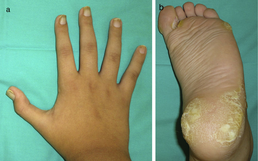
KOH microscopy and culture of nail clippings and oral mucosa ruled out oral thrush and candidal or dermatophytic onychomycosis. Genetic testing confirmed a mutation in the keratin 6a (K6A L469P) in both individuals.
What is your diagnosis?
DiagnosisThe patient and his mother were diagnosed with Pachyonychia Congenita (PC).
DiscussionPC is a rare disorder of keratinization1 described by Muller in 1904 and by Jadassohn and Lewandowsky in 1906. The four main sites of disease involvement include (a) hypertrophic nail dystrophy characterized by yellow-brown thickening of the distal two thirds of the nails with subungual debris resulting in elevation and exaggerated curvature or premature termination of nail plate; (b) painful palmoplantar keratoderma following pressure distribution or involving the entirety of the plantar surface, hyperhidrosis of palms and soles, and fissures at friction sites with occasional blistering and secondary infection; (c) follicular keratoses on the trunk, buttocks and extensor aspects of the extremities that resemble keratosis pilaris, but may coalesce into thick, verrucous plaques; (d) oral leukokeratosis with no inherent malignant potential. PC is also characterized in some cases by natal teeth and presence of pilosebaceous cysts (including steatocystoma and vellus hair cysts).1 Hoarseness due to laryngeal obstruction is very unusual with only a few cases reported.
It is associated with a mutation in one of the five-keratin genes (KRT6A, KRT6B, KRT6C, KRT16, KRT17)2,3 preventing normal filament aggregation, and causing epidermolysis and compensatory hyperkeratosis at these sites. The nail, hair follicle, tongue, palm, and sole are the main sites of constitutive expression of keratins 6, 16, and 17, which corresponds to the distribution of lesions seen in PC. The most commonly mutated gene is KRT6A (≈41–44%), followed by KRT16 (≈25–30%), KRT17 (≈17–24%), KRT6B (≈5–9%) and KRT6C (≈2–3%).3 It was historically divided into two types (PC-1/Jadassohn-Lewandowski and PC-2/Jackson-Lawler); however, its classification is now based on genetic subtypes. This disorder is predominantly inherited in an autosomal dominant pattern with variable expression and a high degree of penetrance, although genetic heterogeneity has been reported.4
Its diagnosis is based on clinical findings; however, genetic confirmation is necessary. Toenail dystrophy is the earliest and most common clinical characteristic of PC in children, which can be present at birth in nearly 40% of PC-affected newborns.5 Genotyping is currently available at no cost on a research basis by registering in the International Pachyonychia Congenita Research Registry (IPCRR, www.pachyonychia.org).
Treatment of PC is notoriously difficult. Treatment of manifestations must focus on grooming of nails and management of pain due to palmoplantar keratoderma, which includes the use of emollients to reduce hyperkeratosis and strategies to limit frictions and trauma of the feet. However, there is no curative treatment for PC yet. When thickened nails are not responsive to topical therapy, extensive surgical removal of nail plate, bed and matrix must be undertaken with subsequent skin grafting. Oral retinoids have been effective for palmoplantar keratodermia in some cases of PC6 and some cases successfully treated with botulinum toxin have been reported.7,8 Moreover, some studies point towards the possibility of neuropathic changes occurring in PC, and therefore neuropathic pain medications could be beneficial in these patients.9 In our case, emollients and keratolytic agents were prescribed, with good disease control.
Please cite this article as: Combalia A, Fustà-Novell X, Estrach T. Onicodistrofia y leucoqueratosis oral en un paciente varón de 15 años. Actas Dermosifiliogr. 2020;111:323–324.


