

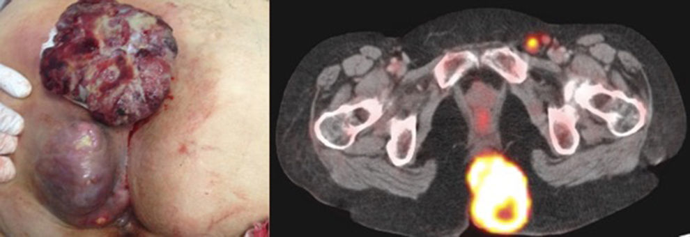
A 57-year-old woman with no past medical history of interest, consulted for 2 large adjacent achromic masses in the sacral region and on the left buttock. One of the lesions was ulcerated. Physical examination revealed 2 erythematous tumors of around 6cm in diameter, with a central area of crusted erosions. Pelvic magnetic resonance imaging showed 2 large, solid heterogeneous masses arising from the dermis in the sacral and left paramedian regions, with no secondary involvement of muscle or bone. The diagnosis on skin biopsy was melanoma in the dermis and subcutaneous tissue, without epidermal involvement. Positron emission tomography-computed tomography showed 2 masses in the left gluteal region, the more cranial of which was exophytic and measured approximately 6.4×3.2cm, with a maximum standardized uptake value (SUVmax) of 28.6. The second mass was in contact with the intergluteal fold and measured approximately 5.0×7.3cm with an SUVmax of 30.3, suggestive of malignancy. Pelvic lymph nodes observed in the left inguinal region were also suggestive of malignancy (Fig. 1).
Please cite this article as: Amo AA, Mínguez ET, Gutzke MM. Melanoma glúteo gigante. Actas Dermosifiliogr. 2017;108:472.


