


Scytalidium spp., recently renamed Neoscytalidium spp., are keratinophilic molds that cause superficial disease (skin, nails) which is indistinguishable from and sometimes occurs concomitantly with dermatophyte infections.1 While infection with these fungi, and in particular Scytalidium dimidiatum, is fairly common in tropical climates, it is rare in Spain. The clinical and microbiological diagnosis of such an infection is a challenge, especially because the growth of these fungi is suppressed by the antimicrobial component of the media routinely used for the isolation of dermatophytes. There is, at present, no effective oral or topical treatment for skin and nail infections caused by Scytalidium.2,3
Antimicrobial photodynamic therapy (PDT) is an emerging treatment for infections. This process involves the application of a photosensitizer that binds to the microorganisms. When excited by an appropriate light source in the presence of oxygen the photosensitizer produces reactive oxygen species that induce cell death, either by apoptosis or necrosis.4 Our group had recently demonstrated the usefulness of methyl-aminolevulinate PDT in the treatment of onychomycosis caused by non-dermatophyte molds5 and yeasts.6
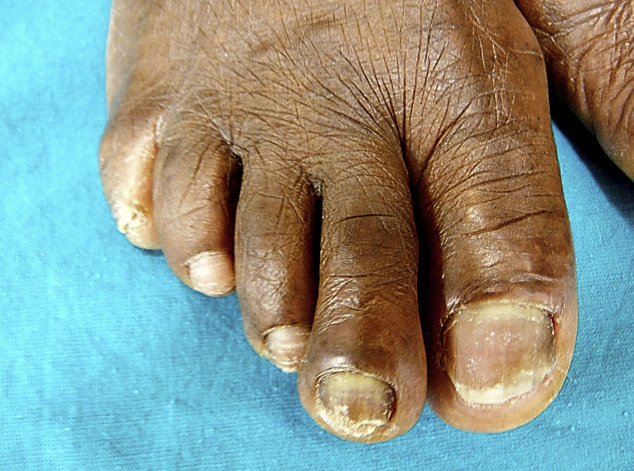
A 61-year-old healthy woman from Cameroon, resident in Spain for more than 5 years, presented with a very long history of dystrophic toenails. She had been diagnosed with onychomycosis caused by Aspergillus spp. and treated with several topical antifungal agents with no improvement. On examination, she presented thickened, opaque nail plates with a yellow or brown discoloration and cracked surfaces (Fig. 1). Significant scaling was observed around the nails, between the toes, and on the soles. Her fingernails were unaffected.

Nail clippings and periungual skin swabs confirmed S. dimidiatum on microscopy and cycloheximide-free agar mycological culture. No dermatophytes were found, but colonies of Aspergillus spp. were seen on Sabouraud plates from 1 of the 2 affected toenails.
Bearing in mind the failure of previous antifungal treatments and the lack of any effective treatment for this infection, and encouraged by our good results in onychomycosis,5,6 proposed this treatment to the patient.
For five days prior to PDT, a combination of 40% urea and 1% bifonazole (Mycospor Onicoset®) ointment was applied every night in occlusion to the nail plates. On the day of treatment, methyl-aminolevulinate (Metvix®) was applied to the nail plates and periungual skin, which were then covered with an occlusive dressing (Tegaderm®) and protected from light for 3h, as previously described.7 When the dressing was removed, the nails were cleaned with 70% ethanol and irradiated using a 635nm light emitting diode lamp (Aktilite®, 37J/cm2). No side effects were observed during or after treatment. The same procedure was repeated 1 week later and every 2 weeks thereafter. Microbiological cultures became negative after the third session. A total of 4 sessions were administered.
Clinical improvement was noticed after 2 months. Four months later, the patient was clinically and microbiologically cured according to the standard criteria (Fig. 2). After 6 months of follow-up, cultures became positive in 1 of the 2 nails, but the nails remained clinically cured and there was no evidence under microscopy of nail penetration.

Infection with S. dimidiatum accounts for under 1% of cases of onychomycosis and seldom responds to amorolfine or terbinafine.2 Recently, an intermittent posaconazole regimen has been proposed to treat superficial S. dimidiatum infection.3 However, all of these drugs are expensive and can have significant adverse effects. Moreover, there is little or no evidence of their effectiveness in this setting.
Fewer than 50 cases of onychomycosis treated with aminolevulinic acid or methyl-aminolevulinate PDT have been reported and most of these were caused by dermatophytes.5–8 Our group obtained good results with methyl-aminolevulinate, using one protocol based on the one previously reported by Piraccini et al.5–7 In localized mycosis, the therapeutic effect of PDT is twofold: the treatment directly kills the fungus and also reinforces the fungicidal effect by stimulating host immune cells, especially neutrophils.4
Although in previous cases5,6 we have successfully used a protocol with a 2-week interval between sessions, in this case we made some modifications. We reduced the interval between the first and second sessions to 1 week in an attempt to reduce the recovery capacity of the fungus. The results of in vitro experiments with dermatophytes suggest that reducing the interval between PDT sessions will improve the fungicidal effect.9
Microbiological diagnosis of onychomycosis is difficult; in fact up to 90% of the cases microbiologically diagnosed the first laboratory result may be negative.10 In our case, however, it is unlikely that the culture results after PDT were false negatives because no antimicrobial substances were present in the medium used. Another problem associated with onychomycosis in general, and Scytalidium infection in particular, is that recurrence is frequent. In this patient we observed microbiological reappearance of the fungus without clinical recurrence. A possible solution for recalcitrant cases might be periodic administration of PDT or a combination of PDT with an antifungal drug over a period of time.
Supported by Grant No. PI120/09 from the Department of Science, Technology and University of the Government of Aragón, Spain and by the research groups B85 and B65 recognized by the Government of Aragón, Spain.


