

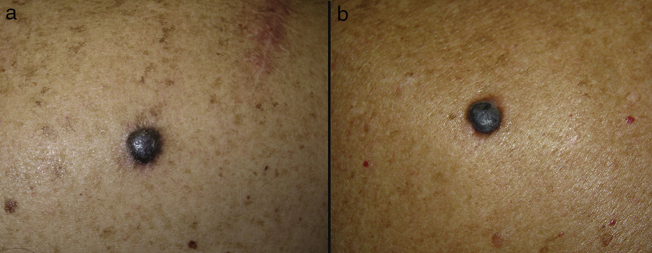
The images show rapidly growing, pigmented nodular lesions in which dermoscopy was very helpful in establishing the diagnosis and, thus, the prognosis (Fig. 1).

A, A pigmented nodular lesion of 10mm diameter that had appeared on the right shoulder of a 54-year-old man 8 months earlier. B, A pigmented nodular lesion of 12mm diameter in the left scapular region of a 67-year-old woman; the patient was uncertain when the lesion had first appeared but reported that it had grown rapidly in recent months.
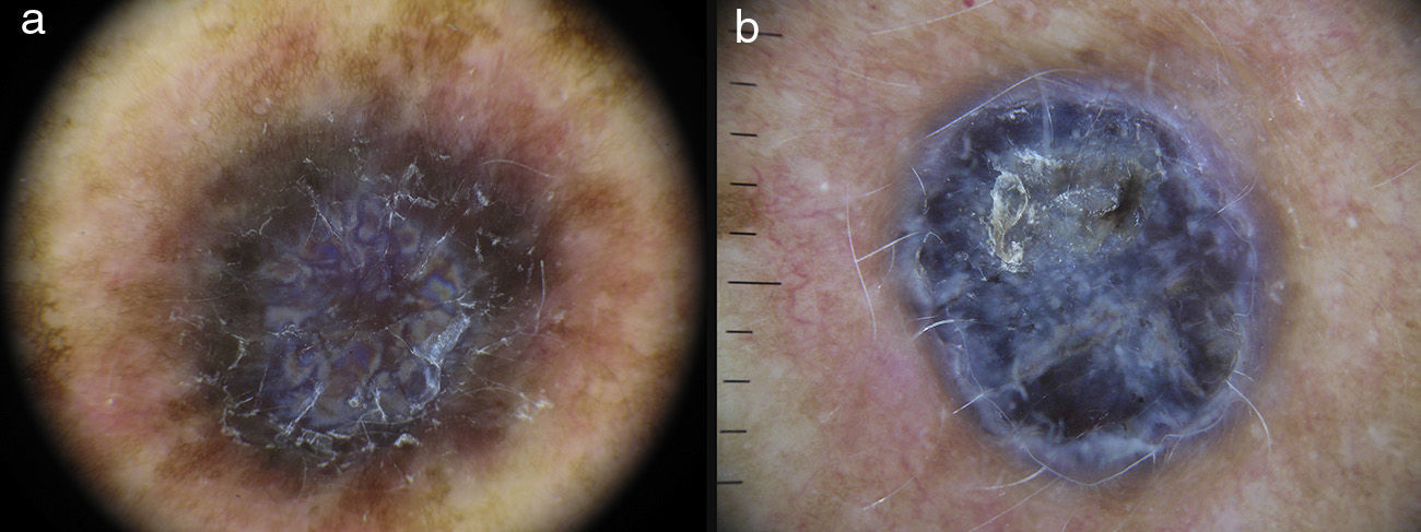
The first case shows a symmetrical lesion with slightly unclear borders, corresponding to a hemosiderotic dermatofibroma. On dermoscopy (Fig. 2A), a polychromic lesion (light and dark brown, red, white, and blue) can be seen with a homogeneous, erythematous-brownish area and a delicate light-brown network peripherally. Attention is drawn to the presence of a rainbow pattern in the central area. This pattern can only be observed with polarized light, with or without contact, and it is thought to be due to an interaction between polarized light and certain skin structures. The finding has been described mainly in Kaposi sarcoma lesions,1 though also in other inflammatory2 and neoplastic3 diseases.

Dermoscopy of hemosiderotic dermatofibroma has also been reported to show the presence of whitish birefringent structures as a result of the passage of polarized light through increased dermal fibrosis,4,5 and the presence of the rainbow phenomenon has been described.2
The second case is of a pigmented nodular lesion that, on dermoscopy is seen to be bichromic (white, blackish-blue), with a scaly crust, foci of ulceration at its superior pole, and pigmented macules at its inferior pole (Fig.2B). The most relevant of these dermoscopic findings were the presence of a blue-gray veil and multiple whitish birefringent areas compatible with chrysalis structures. These signs are suggestive of nodular melanoma.
Chrysalis structures have been described in dermoscopy of melanocytic and nonmelanocytic lesions under polarized light.6
In nodular melanoma, a hypomelanotic/amelanotic lesion is usually seen. In pigmented melanomas, as in our case, the presence of atypical peripheral globules and a whitish-blue veil are the most significant dermoscopic findings. In addition, when chrysalis structures are observed in a melanocytic lesion, spitzoid lesions such as the Spitz nevus, the Reed nevus, and spitzoid melanoma must be included in the differential diagnosis; a malignant lesion must always be excluded, particularly in adult patients.6
In conclusion, we have presented 2 lesions that can be a diagnostic challenge because of their clinical similarities. Dermoscopy played a key role in the investigation of these lesions and revealed different birefringent structures: chrysalis structures in one and a rainbow pattern in the other. The rainbow sign has only recently been described in the literature and its diagnostic value is still to be determined.
Please cite this article as: Padilla-España L, Fernández-Canedo I, Blázquez-Sánchez N. Lesiones nodulares pigmentadas de rápido crecimiento. Actas Dermosifiliogr. 2015;106:505–506.


