


Small-diameter melanomas (≤6mm) are increasingly diagnosed, but their recognition remains challenging because conventional clinical and dermoscopic criteria may be absent or subtle. We investigated their clinical and dermoscopic characteristics and associations with invasiveness.
MethodsThis retrospective, multicenter observational study included 160 histologically confirmed primary cutaneous melanomas measuring ≤6mm, excised between 1998 and 2018. Clinical features were assessed using the ABCDE rule adapted for small melanomas, and global and local dermoscopic characteristics were evaluated for associations with invasiveness.
ResultsOf 160 melanomas, 92 (57.5%) were in situ and 68 (42.5%) were invasive. Only 48 lesions (30%) exhibited at least three ABCDE characteristics, and the overall ABCDE score was not associated with invasiveness. In contrast, invasiveness was significantly associated with multiple suspicious global or local dermoscopic features, more than two colors, peripheral globules, blue-white veil, regression, and polymorphous vessels. Digital dermoscopic surveillance was associated with a higher proportion of in situ melanomas.
ConclusionsA substantial proportion of small-diameter melanomas are already invasive despite limited clinical atypia. Although conventional ABCDE criteria and dermoscopy have diagnostic limitations, specific dermoscopic features and their accumulation may help identify potentially invasive lesions. Digital dermoscopic surveillance may facilitate detection at the in situ stage.
Los diagnósticos de melanomas de pequeño diámetro están aumentando. Su reconocimiento clínico y dermatoscópico puede ser difícil y en un porcentaje relevante de casos ya son melanomas invasores. Este estudio describe las características epidemiológicas, clínicas y dermatoscópicas de melanomas con diámetro ≤6mm y analiza factores relacionados con su invasividad.
Material y métodosEstudio observacional, retrospectivo y multicéntrico de una serie de melanomas con diámetro ≤6mm extirpados entre enero de 1998 y diciembre de 2018. Para su evaluación clínica se empleó la regla ABCDE adaptada a melanomas pequeños y para su evaluación dermatoscópica se consideraron características globales y locales habitualmente descritas en melanoma.
ResultadosSe incluyeron 160 melanomas, 92 (57,5%) in situ y 68 (42,5%) invasores. Solo 48 lesiones (30%) mostraron 3 o más características ABCDE. La puntuación global en la regla ABCDE no se relacionó con la invasividad. La expresividad dermatoscópica se mostró también limitada en muchas de estas lesiones. La acumulación de características dermatoscópicas globales o locales sospechosas en una misma lesión sí se correlacionó con la invasividad, así como la presencia de más de dos colores, de glóbulos periféricos, de regresión gris, de velo azul blanquecino y de vasos polimorfos. El ámbito asistencial de sospecha se asoció con la invasividad destacando el mayor porcentaje de melanomas in situ en pacientes vigilados con dermatoscopia digital.
ConclusionesUn porcentaje elevado de melanomas pequeños ya son invasores. Tanto la regla ABCDE clínica como la dermatoscopia muestran limitaciones para reconocer a muchas de estas lesiones como sospechosas.
Tumor thickness determines the prognosis and associated costs of melanoma.1 Early detection has been promoted by the ABCD mnemonic rule (1985),2 updated to ABCDE in 2004.3 Dermoscopy improves diagnostic sensitivity and specificity.4,5
Early detection has made it possible to identify increasingly thin and small melanomas.6,7 Those measuring ≤6mm account for up to one-third of cases in some series.8–11 The clinical diagnosis of small-diameter melanoma (SDM) can be challenging because these lesions may not meet several or any of the ABCDE criteria,12–14 beginning, by definition, with D (diameter). An adapted ABCDE rule has therefore been proposed for lesions ≤6mm, with D referring to dark (very dark pigmentation).15
Certain melanomas—particularly hypomelanotic or amelanotic,16 nodular,17 and SDM—pose a dermoscopic diagnostic challenge. Individual SDM cases in which dermoscopy was decisive have been reported.18,19 In a series of 206 pigmented lesions measuring<4mm, including 23 melanomas,20 dermoscopy increased diagnostic sensitivity. However, some authors have emphasized the limitations of dermoscopy in recognizing SDMs.11,21–24
Large SDM series with detailed dermoscopic assessments remain scarce. We identified only 13 publications comprising ≥20 cases,11,13,14,23–32 and the largest published series included 123 cases.32
The aim of this study was to analyze 160 SDMs from a multicenter series and correlate their characteristics with invasiveness.
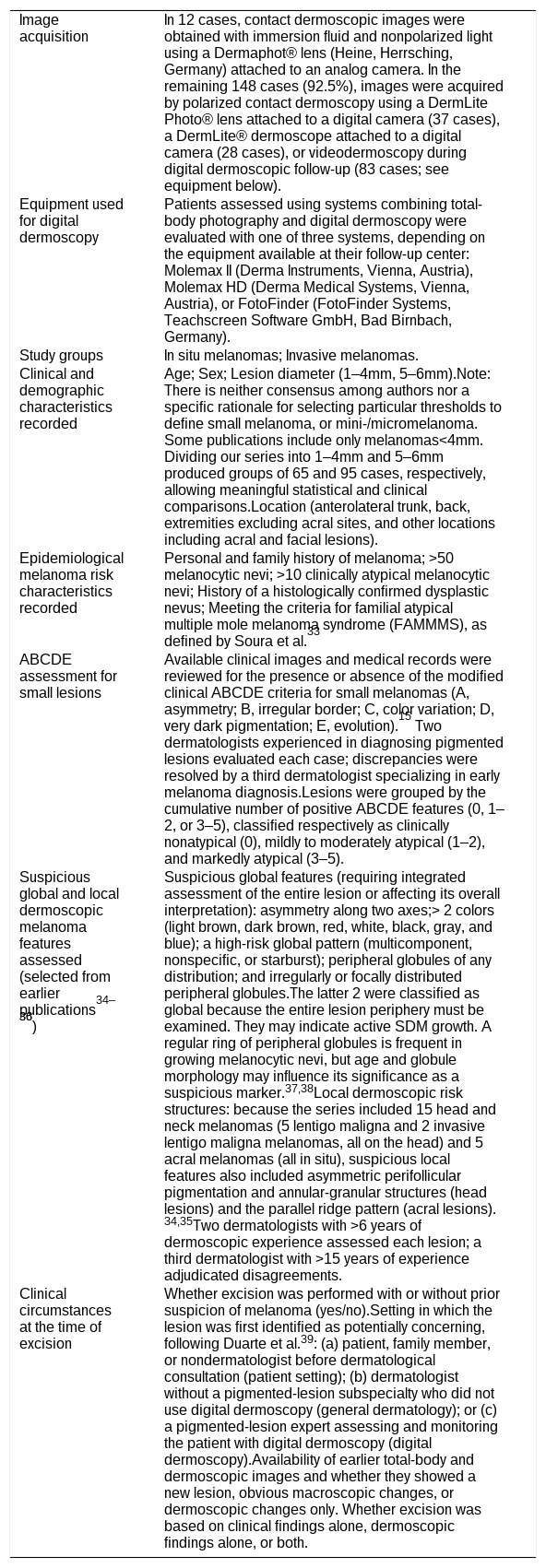
Materials and methodsThis retrospective, observational, descriptive, multicenter study included 160 primary cutaneous melanomas with a clinical diameter ≤6mm (measured on the skin or on the dermoscopic image before excision), for which clinical and dermoscopic images were available. Lesions were excised between 1998 and 2018 in Madrid (Hospital Universitario La Paz, Hospital Universitario La Princesa, Ayuntamiento de Madrid, Instituto Madrileño de Oncología, and Clínica Dermatológica Internacional). All diagnoses were confirmed histologically and reviewed by two expert dermatopathologists. Methodological details are presented in Table 1.
Study methods, study groups, and variables assessed.
| Image acquisition | In 12 cases, contact dermoscopic images were obtained with immersion fluid and nonpolarized light using a Dermaphot® lens (Heine, Herrsching, Germany) attached to an analog camera. In the remaining 148 cases (92.5%), images were acquired by polarized contact dermoscopy using a DermLite Photo® lens attached to a digital camera (37 cases), a DermLite® dermoscope attached to a digital camera (28 cases), or videodermoscopy during digital dermoscopic follow-up (83 cases; see equipment below). |
| Equipment used for digital dermoscopy | Patients assessed using systems combining total-body photography and digital dermoscopy were evaluated with one of three systems, depending on the equipment available at their follow-up center:Molemax II (Derma Instruments, Vienna, Austria), Molemax HD (Derma Medical Systems, Vienna, Austria), or FotoFinder (FotoFinder Systems, Teachscreen Software GmbH, Bad Birnbach, Germany). |
| Study groups | In situ melanomas; Invasive melanomas. |
| Clinical and demographic characteristics recorded | Age; Sex; Lesion diameter (1–4mm, 5–6mm).Note: There is neither consensus among authors nor a specific rationale for selecting particular thresholds to define small melanoma, or mini-/micromelanoma. Some publications include only melanomas<4mm. Dividing our series into 1–4mm and 5–6mm produced groups of 65 and 95 cases, respectively, allowing meaningful statistical and clinical comparisons.Location (anterolateral trunk, back, extremities excluding acral sites, and other locations including acral and facial lesions). |
| Epidemiological melanoma risk characteristics recorded | Personal and family history of melanoma; >50 melanocytic nevi; >10 clinically atypical melanocytic nevi; History of a histologically confirmed dysplastic nevus; Meeting the criteria for familial atypical multiple mole melanoma syndrome (FAMMMS), as defined by Soura et al.33 |
| ABCDE assessment for small lesions | Available clinical images and medical records were reviewed for the presence or absence of the modified clinical ABCDE criteria for small melanomas (A, asymmetry; B, irregular border; C, color variation; D, very dark pigmentation; E, evolution).15 Two dermatologists experienced in diagnosing pigmented lesions evaluated each case; discrepancies were resolved by a third dermatologist specializing in early melanoma diagnosis.Lesions were grouped by the cumulative number of positive ABCDE features (0, 1–2, or 3–5), classified respectively as clinically nonatypical (0), mildly to moderately atypical (1–2), and markedly atypical (3–5). |
| Suspicious global and local dermoscopic melanoma features assessed (selected from earlier publications34–36) | Suspicious global features (requiring integrated assessment of the entire lesion or affecting its overall interpretation): asymmetry along two axes;> 2 colors (light brown, dark brown, red, white, black, gray, and blue); a high-risk global pattern (multicomponent, nonspecific, or starburst); peripheral globules of any distribution; and irregularly or focally distributed peripheral globules.The latter 2 were classified as global because the entire lesion periphery must be examined. They may indicate active SDM growth. A regular ring of peripheral globules is frequent in growing melanocytic nevi, but age and globule morphology may influence its significance as a suspicious marker.37,38Local dermoscopic risk structures: because the series included 15 head and neck melanomas (5 lentigo maligna and 2 invasive lentigo maligna melanomas, all on the head) and 5 acral melanomas (all in situ), suspicious local features also included asymmetric perifollicular pigmentation and annular-granular structures (head lesions) and the parallel ridge pattern (acral lesions).34,35Two dermatologists with >6 years of dermoscopic experience assessed each lesion; a third dermatologist with >15 years of experience adjudicated disagreements. |
| Clinical circumstances at the time of excision | Whether excision was performed with or without prior suspicion of melanoma (yes/no).Setting in which the lesion was first identified as potentially concerning, following Duarte et al.39: (a) patient, family member, or nondermatologist before dermatological consultation (patient setting); (b) dermatologist without a pigmented-lesion subspecialty who did not use digital dermoscopy (general dermatology); or (c) a pigmented-lesion expert assessing and monitoring the patient with digital dermoscopy (digital dermoscopy).Availability of earlier total-body and dermoscopic images and whether they showed a new lesion, obvious macroscopic changes, or dermoscopic changes only. Whether excision was based on clinical findings alone, dermoscopic findings alone, or both. |
We assessed possible associations between lesion invasiveness (in situ vs invasive) and 3 groups of variables: patient history and characteristics; clinical and dermoscopic tumor features; and the clinical circumstances in which excision was indicated.
Chi-square or Fisher exact tests were used as appropriate, and odds ratios were calculated for selected variables. Statistical significance was set at P<.05. The study was approved by the Research Ethics Committee of Hospital Universitario La Princesa (No. 2760, minutes 06/2016).
ResultsWe analyzed a total of 160 cases in 151 patients (mean age, 47 years; range, 14–84 years): 144 patients had 1 melanoma, 5 had 2, and 2 had 3. Overall, 57.5% were in situ (including seven lentigo maligna melanomas) and 42.5% were invasive (median thickness, 0.43mm; interquartile range, 0.3–0.6mm). Only three lesions exceeded a Breslow thickness of 1mm (2 nodular melanomas measuring 3mm in diameter, with thicknesses of 1.1 and 2.5mm, respectively, and 1 superficial spreading melanoma with a Breslow thickness of 1.2mm). None showed ulceration, and only 5 showed mitoses, including the three thickest lesions (sentinel lymph node biopsies were negative; all three patients remain alive and recurrence-free after ≥10 years of follow-up).
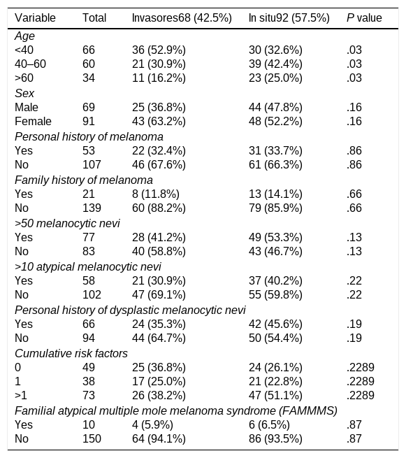
Table 2 details the demographic and epidemiological characteristics of the patients. The most common factor associated with increased melanoma risk or difficulties in surveillance was the presence of >50 nevi (48.1% of cases). Overall, 30.6% of patients had none of these factors. Only age at diagnosis was associated with invasiveness, with invasive tumors more frequent in patients <40 years of age.
Demographic and epidemiological characteristics of the patients. Comparison of in situ and invasive melanomas (N=160).
| Variable | Total | Invasores68 (42.5%) | In situ92 (57.5%) | P value |
|---|---|---|---|---|
| Age | ||||
| <40 | 66 | 36 (52.9%) | 30 (32.6%) | .03 |
| 40–60 | 60 | 21 (30.9%) | 39 (42.4%) | .03 |
| >60 | 34 | 11 (16.2%) | 23 (25.0%) | .03 |
| Sex | ||||
| Male | 69 | 25 (36.8%) | 44 (47.8%) | .16 |
| Female | 91 | 43 (63.2%) | 48 (52.2%) | .16 |
| Personal history of melanoma | ||||
| Yes | 53 | 22 (32.4%) | 31 (33.7%) | .86 |
| No | 107 | 46 (67.6%) | 61 (66.3%) | .86 |
| Family history of melanoma | ||||
| Yes | 21 | 8 (11.8%) | 13 (14.1%) | .66 |
| No | 139 | 60 (88.2%) | 79 (85.9%) | .66 |
| >50 melanocytic nevi | ||||
| Yes | 77 | 28 (41.2%) | 49 (53.3%) | .13 |
| No | 83 | 40 (58.8%) | 43 (46.7%) | .13 |
| >10 atypical melanocytic nevi | ||||
| Yes | 58 | 21 (30.9%) | 37 (40.2%) | .22 |
| No | 102 | 47 (69.1%) | 55 (59.8%) | .22 |
| Personal history of dysplastic melanocytic nevi | ||||
| Yes | 66 | 24 (35.3%) | 42 (45.6%) | .19 |
| No | 94 | 44 (64.7%) | 50 (54.4%) | .19 |
| Cumulative risk factors | ||||
| 0 | 49 | 25 (36.8%) | 24 (26.1%) | .2289 |
| 1 | 38 | 17 (25.0%) | 21 (22.8%) | .2289 |
| >1 | 73 | 26 (38.2%) | 47 (51.1%) | .2289 |
| Familial atypical multiple mole melanoma syndrome (FAMMMS) | ||||
| Yes | 10 | 4 (5.9%) | 6 (6.5%) | .87 |
| No | 150 | 64 (94.1%) | 86 (93.5%) | .87 |
Rightmost column: Chi-square P values (P<.05 shown in bold).
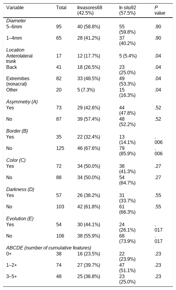
The clinical characteristics of melanomas are summarized in Table 3. Notably, 65 tumors measured ≤4mm (minimelanomas), of which 43% were already invasive. The extremities accounted for 51% of cases (excluding acral lesions), most frequently the lower extremities (34.4% of the series, not separately shown in the table). Of these 55 cases on the lower extremities, 75% occurred in women.
Clinical characteristics of melanomas. Comparison of in situ and invasive melanomas (N=160).
| Variable | Total | Invasores68 (42.5%) | In situ92 (57.5%) | P value |
|---|---|---|---|---|
| Diameter | ||||
| 5–6mm | 95 | 40 (58.8%) | 55 (59.8%) | .90 |
| 1–4mm | 65 | 28 (41.2%) | 37 (40.2%) | .90 |
| Location | ||||
| Anterolateral trunk | 17 | 12 (17.7%) | 5 (5.4%) | .04 |
| Back | 41 | 18 (26.5%) | 23 (25.0%) | .04 |
| Extremities (nonacral) | 82 | 33 (48.5%) | 49 (53.3%) | .04 |
| Other | 20 | 5 (7.3%) | 15 (16.3%) | .04 |
| Asymmetry (A) | ||||
| Yes | 73 | 29 (42.6%) | 44 (47.8%) | .52 |
| No | 87 | 39 (57.4%) | 48 (52.2%) | .52 |
| Border (B) | ||||
| Yes | 35 | 22 (32.4%) | 13 (14.1%) | .006 |
| No | 125 | 46 (67.6%) | 79 (85.9%) | .006 |
| Color (C) | ||||
| Yes | 72 | 34 (50.0%) | 38 (41.3%) | .27 |
| No | 88 | 34 (50.0%) | 54 (64.7%) | .27 |
| Darkness (D) | ||||
| Yes | 57 | 26 (38.2%) | 31 (33.7%) | .55 |
| No | 103 | 42 (61.8%) | 61 (66.3%) | .55 |
| Evolution (E) | ||||
| Yes | 54 | 30 (44.1%) | 24 (26.1%) | .017 |
| No | 106 | 38 (55.9%) | 68 (73.9%) | .017 |
| ABCDE (number of cumulative features) | ||||
| 0+ | 38 | 16 (23.5%) | 22 (23.9%) | .23 |
| 1–2+ | 74 | 27 (39.7%) | 47 (51.1%) | .23 |
| 3–5+ | 48 | 25 (36.8%) | 23 (25.0%) | .23 |
A-L: anterolateral.
Rightmost column: chi-square P values (P<.05 shown in bold).
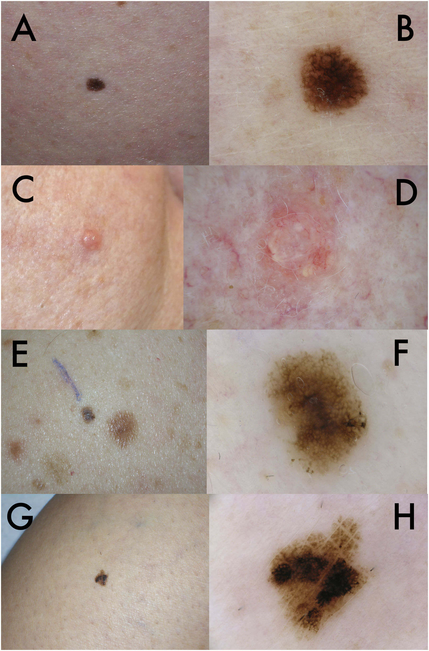
Regarding the number of ABCDE characteristics (Table 3), 24% had none (clinically nonatypical; Fig. 1A and C), 46% had 1 or 2 (mild-to-moderate atypia; Fig. 1E), and 30% had ≥3 (marked clinical atypia; Fig. 1G). Invasive melanomas were more common on the anterolateral trunk. Irregular borders (B) and changes observed by the patient (E) were significantly associated with invasiveness.
Clinical images (A, C, E, and G) and corresponding dermoscopic images (B, D, F, and H) of 4 small melanomas. (A) In situ melanoma on the thigh of a 49-year-old woman, with growth unnoticed by the patient and a clinically nonatypical appearance. (B) Poorly structured pigment network as the only relevant dermoscopic finding. (C) Nodular melanoma presenting as a 3-mm pink facial papule in a 65-year-old woman, meeting no ABCD criteria and measuring 2.5mm in thickness. (D) Focal polymorphous vessels on dermoscopy. (E) Superficial spreading melanoma on the back of a 33-year-old man, 0.5mm thick, with minimal clinical atypia and growth detected by total-body photographs. (F) Clearly suspicious dermoscopy showing asymmetry along two axes, an atypical network, and focal peripheral globules. (G) In situ melanoma on the leg of a 76-year-old woman with marked clinical atypia. (H) Highly suspicious dermoscopy, with marked asymmetry, irregular pigmentation, and areas of prominent atypical network.
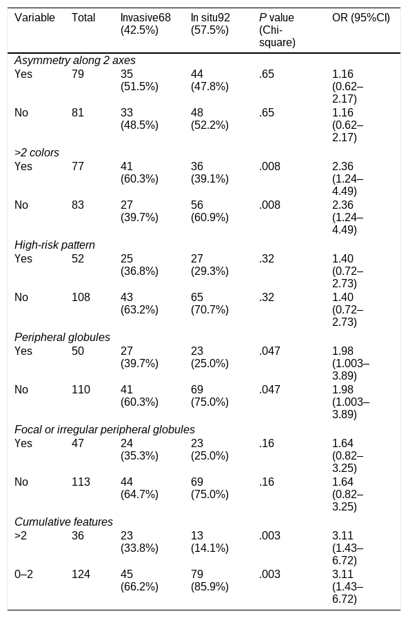
No suspicious global dermoscopic feature occurred in >50% of cases (Table 4). The most common were asymmetry along two axes (49%) and >2 colors (48%). Invasiveness was significantly associated with >2 colors, peripheral globules irrespective of their distribution (Fig. 1F), and the accumulation of several suspicious global features within one lesion. Notably, all three melanomas displaying regularly distributed peripheral globules in a ring were invasive and occurred in patients aged 36, 39, and 54 years (Breslow thicknesses, 0.35, 0.4, and 0.3mm).
Global dermoscopic risk features. Comparison of in situ and invasive melanomas (N=160).
| Variable | Total | Invasive68 (42.5%) | In situ92 (57.5%) | P value (Chi-square) | OR (95%CI) |
|---|---|---|---|---|---|
| Asymmetry along 2 axes | |||||
| Yes | 79 | 35 (51.5%) | 44 (47.8%) | .65 | 1.16 (0.62–2.17) |
| No | 81 | 33 (48.5%) | 48 (52.2%) | .65 | 1.16 (0.62–2.17) |
| >2 colors | |||||
| Yes | 77 | 41 (60.3%) | 36 (39.1%) | .008 | 2.36 (1.24–4.49) |
| No | 83 | 27 (39.7%) | 56 (60.9%) | .008 | 2.36 (1.24–4.49) |
| High-risk pattern | |||||
| Yes | 52 | 25 (36.8%) | 27 (29.3%) | .32 | 1.40 (0.72–2.73) |
| No | 108 | 43 (63.2%) | 65 (70.7%) | .32 | 1.40 (0.72–2.73) |
| Peripheral globules | |||||
| Yes | 50 | 27 (39.7%) | 23 (25.0%) | .047 | 1.98 (1.003–3.89) |
| No | 110 | 41 (60.3%) | 69 (75.0%) | .047 | 1.98 (1.003–3.89) |
| Focal or irregular peripheral globules | |||||
| Yes | 47 | 24 (35.3%) | 23 (25.0%) | .16 | 1.64 (0.82–3.25) |
| No | 113 | 44 (64.7%) | 69 (75.0%) | .16 | 1.64 (0.82–3.25) |
| Cumulative features | |||||
| >2 | 36 | 23 (33.8%) | 13 (14.1%) | .003 | 3.11 (1.43–6.72) |
| 0–2 | 124 | 45 (66.2%) | 79 (85.9%) | .003 | 3.11 (1.43–6.72) |
Penultimate column: chi-square P values (P<.05 shown in bold).
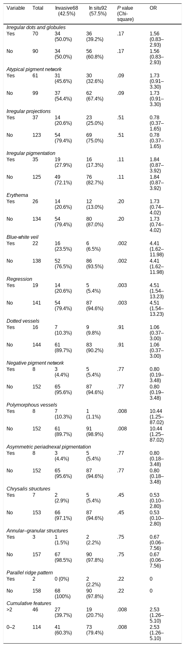
Among local features (Table 5), irregular dots/globules (44%) and an atypical pigment network (38%) were most frequent (Fig. 1B, F, and H). A blue-white veil, regression, and polymorphous vessels (Fig. 1D), together with an increasing accumulation of suspicious local features in the same lesion, were significantly associated with invasiveness.
Local dermoscopic risk features. Comparison of in situ and invasive melanomas (N=160).
| Variable | Total | Invasive68 (42.5%) | In situ92 (57.5%) | P value (Chi-square) | OR |
|---|---|---|---|---|---|
| Irregular dots and globules | |||||
| Yes | 70 | 34 (50.0%) | 36 (39.2%) | .17 | 1.56 (0.83–2.93) |
| No | 90 | 34 (50.0%) | 56 (60.8%) | .17 | 1.56 (0.83–2.93) |
| Atypical pigment network | |||||
| Yes | 61 | 31 (45.6%) | 30 (32.6%) | .09 | 1.73 (0.91–3.30) |
| No | 99 | 37 (54.4%) | 62 (67.4%) | .09 | 1.73 (0.91–3.30) |
| Irregular projections | |||||
| Yes | 37 | 14 (20.6%) | 23 (25.0%) | .51 | 0.78 (0.37–1.65) |
| No | 123 | 54 (79.4%) | 69 (75.0%) | .51 | 0.78 (0.37–1.65) |
| Irregular pigmentation | |||||
| Yes | 35 | 19 (27.9%) | 16 (17.3%) | .11 | 1.84 (0.87–3.92) |
| No | 125 | 49 (72.1%) | 76 (82.7%) | .11 | 1.84 (0.87–3.92) |
| Erythema | |||||
| Yes | 26 | 14 (20.6%) | 12 (13.0%) | .20 | 1.73 (0.74–4.02) |
| No | 134 | 54 (79.4%) | 80 (87.0%) | .20 | 1.73 (0.74–4.02) |
| Blue-white veil | |||||
| Yes | 22 | 16 (23.5%) | 6 (6.5%) | .002 | 4.41 (1.62–11.98) |
| No | 138 | 52 (76.5%) | 86 (93.5%) | .002 | 4.41 (1.62–11.98) |
| Regression | |||||
| Yes | 19 | 14 (20.6%) | 5 (5.4%) | .003 | 4.51 (1.54–13.23) |
| No | 141 | 54 (79.4%) | 87 (94.6%) | .003 | 4.51 (1.54–13.23) |
| Dotted vessels | |||||
| Yes | 16 | 7 (10.3%) | 9 (9.8%) | .91 | 1.06 (0.37–3.00) |
| No | 144 | 61 (89.7%) | 83 (90.2%) | .91 | 1.06 (0.37–3.00) |
| Negative pigment network | |||||
| Yes | 8 | 3 (4.4%) | 5 (5.4%) | .77 | 0.80 (0.19–3.48) |
| No | 152 | 65 (95.6%) | 87 (94.6%) | .77 | 0.80 (0.19–3.48) |
| Polymorphous vessels | |||||
| Yes | 8 | 7 (10.3%) | 1 (1.1%) | .008 | 10.44 (1.25–87.02) |
| No | 152 | 61 (89.7%) | 91 (98.9%) | .008 | 10.44 (1.25–87.02) |
| Asymmetric periadnexal pigmentation | |||||
| Yes | 8 | 3 (4.4%) | 5 (5.4%) | .77 | 0.80 (0.18–3.48) |
| No | 152 | 65 (95.6%) | 87 (94.6%) | .77 | 0.80 (0.18–3.48) |
| Chrysalis structures | |||||
| Yes | 7 | 2 (2.9%) | 5 (5.4%) | .45 | 0.53 (0.10–2.80) |
| No | 153 | 66 (97.1%) | 87 (94.6%) | .45 | 0.53 (0.10–2.80) |
| Annular–granular structures | |||||
| Yes | 3 | 1 (1.5%) | 2 (2.2%) | .75 | 0.67 (0.06–7.56) |
| No | 157 | 67 (98.5%) | 90 (97.8%) | .75 | 0.67 (0.06–7.56) |
| Parallel ridge pattern | |||||
| Yes | 2 | 0 (0%) | 2 (2.2%) | .22 | 0 |
| No | 158 | 68 (100%) | 90 (97.8%) | .22 | 0 |
| Cumulative features | |||||
| >2 | 46 | 27 (39.7%) | 19 (20.7%) | .008 | 2.53 (1.26–5.10) |
| 0–2 | 114 | 41 (60.3%) | 73 (79.4%) | .008 | 2.53 (1.26–5.10) |
Penultimate column: chi-square P values (P<.05 shown in bold).
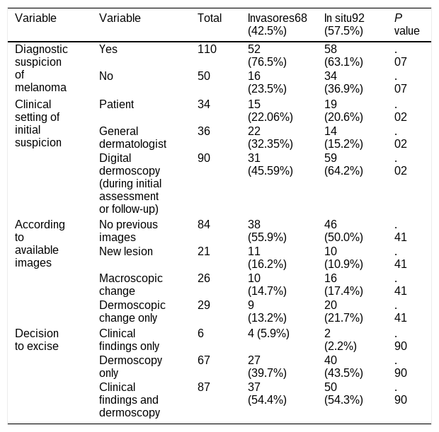
Finally, Table 6 describes the clinical circumstances. Of note, 56% of cases were detected in a specialized pigmented-lesion clinic equipped with digital dermoscopy. The clinical setting was significantly associated with invasiveness, with in situ melanomas occurring more frequently among patients monitored using digital dermoscopy.
Clinical circumstances when excision was decided. Comparison of in situ and invasive melanomas (N=160).
| Variable | Variable | Total | Invasores68 (42.5%) | In situ92 (57.5%) | P value |
|---|---|---|---|---|---|
| Diagnostic suspicion of melanoma | Yes | 110 | 52 (76.5%) | 58 (63.1%) | .07 |
| No | 50 | 16 (23.5%) | 34 (36.9%) | .07 | |
| Clinical setting of initial suspicion | Patient | 34 | 15 (22.06%) | 19 (20.6%) | .02 |
| General dermatologist | 36 | 22 (32.35%) | 14 (15.2%) | .02 | |
| Digital dermoscopy (during initial assessment or follow-up) | 90 | 31 (45.59%) | 59 (64.2%) | .02 | |
| According to available images | No previous images | 84 | 38 (55.9%) | 46 (50.0%) | .41 |
| New lesion | 21 | 11 (16.2%) | 10 (10.9%) | .41 | |
| Macroscopic change | 26 | 10 (14.7%) | 16 (17.4%) | .41 | |
| Dermoscopic change only | 29 | 9 (13.2%) | 20 (21.7%) | .41 | |
| Decision to excise | Clinical findings only | 6 | 4 (5.9%) | 2 (2.2%) | .90 |
| Dermoscopy only | 67 | 27 (39.7%) | 40 (43.5%) | .90 | |
| Clinical findings and dermoscopy | 87 | 37 (54.4%) | 50 (54.3%) | .90 | |
This study analyzes the largest published series to date of SDMs evaluated by dermoscopy and confirms their limited clinical and dermoscopic expression, as reported in several former series.11,14,21,23,24,29,31 Comparison between studies is difficult because of methodological differences. We therefore focused this discussion on published series with ≥20 cases and sufficiently detailed descriptions of dermoscopic findings.11,13,14,23–32
Particularly noteworthy is that 42.5% of all our SDMs (and 43.1% of the 65 lesions measuring ≤4mm) were already invasive. This proportion varies widely across series11,13,14,23–32,40–42: among those providing detailed dermoscopic assessments, it ranges from 4%30 to 72.4%.27 Some of these invasive SDMs are metastatic,8,43 with nodular melanoma predominating among them43; this subtype often grows rapidly.6
The absence of a significant correlation between SDM diameter and invasiveness in our series (as also observed in series of larger melanomas44,45) suggests that some melanomas are biologically more aggressive from the outset, whereas others are destined to follow a more indolent course as in situ lesions until they reach diameters frequently exceeding 6mm.
Our findings highlight the limitations of the ABCDE rule for recognizing many SDMs, consistent with previous reports.13,14,32 The absence of an association between invasiveness and ABCDE-defined clinical atypia in this and other series39,46 also illustrates the limitations of clinical examination in determining whether a small suspicious lesion may already be invasive. Recognition of nodular SDMs poses a particular challenge because they commonly meet only the E criterion or none of the clinical ABCDE criteria.47 Indeed, one nodular melanoma in our series met none (Fig. 1C), and the other met only E. In our opinion, shared by other melanoma early-detection experts,48,49 every small, equivocal, palpable melanocytic lesion warrants particular caution and should preferably be excised in the absence of evidence confirming benignity.
The association of irregular borders (B) with invasiveness should be interpreted cautiously because nodular melanomas (usually more invasive and with regular borders)47 were probably underrepresented in our series: some may have been excised without specific suspicion of melanoma or photographic documentation. Conversely, the association of evolution (E) with invasiveness is consistent with findings in series not restricted by lesion size.39,50 Thus, a small, equivocal melanocytic lesion warrants particular caution when the patient reports recent changes.
SDMs predominate in younger adults: the mean age of our cohort (47.1 years) is approximately 10 years lower than that of patients with melanoma in our setting irrespective of lesion diameter.51,52 Their frequent occurrence on the extremities, particularly on women's legs, is also notable and has been described by other authors.13,53,54 We believe this finding indicates the patients in whom melanoma is most likely to be detected and excised while still small.
Although dermoscopic features are often subtle, some SDMs can be recognized readily by dermoscopy (Fig. 1F and H), which can also reveal key suspicious features in clinically unremarkable lesions (Fig. 1C and D).
The dermoscopic appearance of SDMs varies considerably across studies.11,13,14,23–32 Common suspicious global findings include those assessed here—asymmetry, multiple colors, and a high-risk global pattern (multicomponent, nonspecific, or spitzoid)—although a substantial proportion of tumors have only one or none of these signs,11,13,14,23–32 as in our study and in contrast to larger melanomas. Among local structures, irregular dots/globules and atypical pigment network were the most common in our series and are also frequent in other SDM series.11,13,14,23–32 Other features found relatively often in some but not all series include irregular pigmentation, irregular blotches, radial streaming, pseudopods, blue-white veil, and regression structures. Negative pigment network, shiny white lines/chrysalis structures, and atypical vascularization are less common. The low frequency of shiny white lines/chrysalis structures (4.4%) in our series does not appear attributable to nonpolarized dermoscopy, because only 12 cases (7.5%) were imaged without polarized light. We did not assess 2 dermoscopic structures described after our study design and case review began: angulated lines/polygons and superficial linear skin markings. Although one series of 26 cases found these in 46.2% and 11.5% of cases, respectively,26 other more recent and/or much larger series either detected none29 or very few.11,31
Quantifying the frequency of each dermoscopic structure helps establish what clinicians should look for and what can reasonably be expected in SDMs.11,13,14,23–32 However, assessing the tendency for suspicious global and local findings to accumulate, as we also did, more clearly reveals the diagnostic complexity. In our series, 49% of lesions displayed ≤1 global criterion and 71% displayed ≤2 local criteria, proportions similar to those seen in many atypical nevi. This explains why published algorithms perform less well for SDMs than for larger melanomas11,23,24,29,31,32 and confirms that most SDMs are difficult to detect dermoscopically, although views differ.20,25
Clinical atypia was not associated with invasiveness in our series, whereas dermoscopic atypia was. Specifically, invasiveness was associated with the accumulation of suspicious global or local dermoscopic melanoma features and with individual findings such as >2 colors, peripheral globules of any distribution pattern, blue-white veil, regression, and polymorphous vessels. Other authors have reported associations with invasiveness in SDMs for atypical vascular patterns, blue-gray structures, chrysalis structures, and negative pigment networks.11,31 When a small, equivocal nevus shows these signs, we recommend excision.
A striking finding was that all 3 melanomas with regularly arranged peripheral globules (in a ring) were invasive. Although a ring of peripheral globules is often seen in growing benign nevi, caution is necessary if the globules are irregular in morphology, color, or size; if the lesion displays any other central atypical feature; or if the finding occurs in middle-aged or older adults.37,38 Our study shows that, although uncommon, a regular ring of peripheral globules may accompany invasive SDM.
Of the melanomas in this series, 68.8% were excised with melanoma considered in the differential diagnosis. Dermoscopy was decisive in the decision to excise 96.3% of cases (in 87 cases together with clinical information), despite minimal clinical atypia and limited dermoscopic features in many lesions. In some instances, excision may have been based on evidence of evolution—the age at which a nevus appeared or changed—even in the absence of clear malignant features.55
Surveillance with total-body photography and digital dermoscopy facilitates detection of new or changing lesions.11,13,23,28,40,56,57 In our series, most melanomas detected in this way were in situ, and the clinical setting was associated with invasiveness. Clearly, more intensive surveillance11,13,23,28,40 makes it easier to identify new or unstable lesions while still small and noninvasive. Histologic diagnosis of in situ or thin melanoma leads to smaller final scars, but whether it reduces overall melanoma mortality remains debatable.58
Regarding potential overdiagnosis of melanomas that are histologically genuine but biologically harmless,58,59 we believe that restricting surgery to lesions>6mm, as proposed by some authors,58 is dangerous: many SDMs are already invasive and some metastasize.8,43 A more prudent strategy is to excise small suspicious lesions showing signs of invasiveness and consider monitoring selected lesions without such signs. Dermoscopy appears helpful in this task, as suggested by our results and those of other studies,11,31 provided it is not used to monitor palpable or nodular lesions of uncertain nature.48,49 Although digital dermoscopy may contribute in part to overdiagnosis,58 we believe it can also help address it,59 by avoiding excision of most flat atypical nevi that remain stable or grow slowly without developing further atypia.
Some SDM series evaluated by dermoscopy have also used reflectance confocal microscopy.13,23,25,30 This technique unquestionably increases diagnostic specificity.60 Moreover, recent research61 suggests that it may provide information about biological aggressiveness, potentially reducing overdiagnosis.62 It remains to be seen what artificial intelligence-based diagnostic support systems can offer,63 both for detecting SDMs and for identifying those that are biologically more aggressive and should always be excised. Recognizing the strengths and weaknesses of melanoma diagnostic strategies64 is essential to optimize outcomes for our patients.
LimitationsThe main limitations are the retrospective design, heterogeneity of imaging equipment, and requirement for photographic documentation, which may have excluded unsuspected SDMs, particularly nodular melanomas.
ConclusionsMany SDMs are already invasive. They occur predominantly in younger adults and on the legs of women. The adapted clinical ABCDE rule (with D meaning dark pigmentation) has limitations in recognizing them or predicting invasiveness. SDMs are also challenging to detect by dermoscopy, although this technique reveals certain useful clues to potentially invasive lesions. More than half of the SDMs in this series were suspected among patients undergoing digital dermoscopic surveillance, with a significantly higher proportion of in situ lesions. Further studies and strategies are needed to facilitate early diagnosis while minimizing overdiagnosis.
Conflict of interestThe authors declare that they have no conflict of interest.