


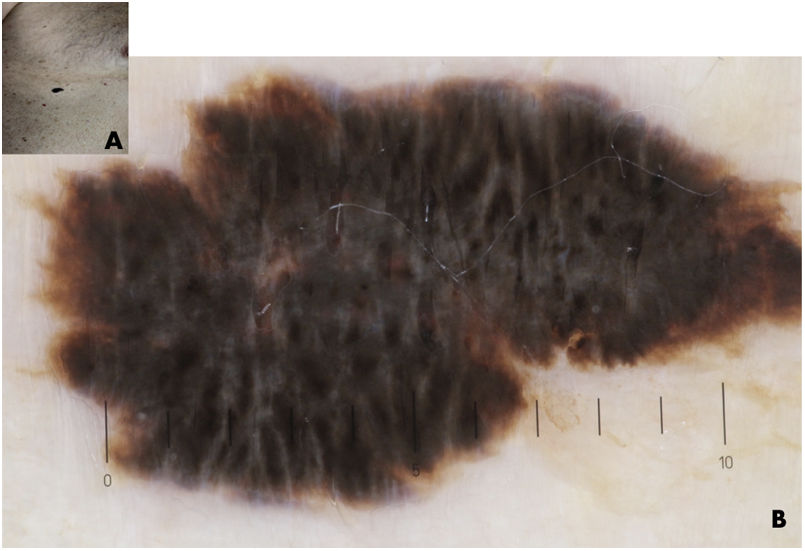
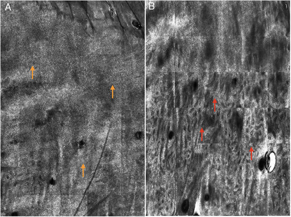
Reflectance confocal microscopy (RCM) is a not invasive technique for in vivo imaging of the skin that uses a near-infrared laser beam of 830 nm. Nowadays, RCM imaging is mainly used for melanoma and non-melanoma diagnosis, particularly in those challenging cases in which dermatoscopy is not so helpful. A 74 y/o men presented to our department with a heavily pigmented lesion on the right abdomen (Fig. 1) appeared for about a year and grew slowly. On dermatoscopy the lesion did not show elements that made it define its exact nature (melanocytic or non-melanocytic). The heavily black pigmentation of the lesion, the presence of a central veil-like area (usually observed in malignant melanocytic lesions) and finally the tendency to increase the lesion in size, have made the dermatoscopic examination alone not sufficient, pushing us to use confocal laser microscopy. RCM (Fig. 2) highlighted the presence of a physiologic granular-spinous layer without disarrangement, atypical cells or malignant spreading. Deeper the acquisition was characterized by bulbous projection of the dermo-epidermal junction with findings were compatible with diagnosis of a heavily pigmented seborrheic keratosis (SK) also known as melanoacanthoma, an infrequent variant of SK that is clinically notable for its dark pigmentation. Few data regarding the utility of RCM on diagnosis of melanoacanthoma are detectable in literature. Shahriari et al. (2016) published a case of a 51 y/o women affected by melanoacanthoma in which the acquisition, being characterized by hyperplastic epidermis with numerous tangled dendritic cells, could not rule out an atypical melanocytic lesion. Our experience underlines that RCM is a useful tool for melanoacanthoma diagnosis.
Please cite this article as: Mazzeo M, Manfreda V, Botti E, Bianchi L. Papel de la microscopia confocal de reflectancia en el diagnóstico del melanoacantoma. Actas Dermosifiliogr. 2021. https://doi.org/10.1016/j.ad.2019.07.025


