


A 68-year-old hypertensive woman consulted about an asymptomatic lesion of 1 year's duration at her annual checkup. Her last check up had been 2 years earlier, and there had been no remarkable findings.
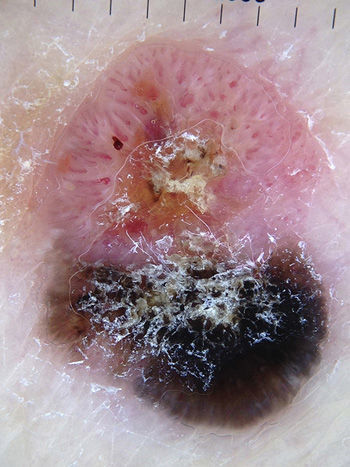
Physical ExaminationThe physical examination revealed a slightly indurated tumor measuring 2cm on the left buttock. The lesion had 2 components: a pink erythematous component with a vascular appearance and a white-yellowish scale-crust, and a second component with brown pigmentation (Fig. 1). Dermoscopic examination showed polymorphous (dotted and hairpin) vessels surrounded by a whitish halo in the vascularized component and brown pigment with peripheral projections that did not radiate from the center of the lesion or form a specific dermoscopic pattern in the pigmented component (Fig. 2). An excisional biopsy was performed to check for melanoma and pigmented basal cell carcinoma (BCC).
Histopathology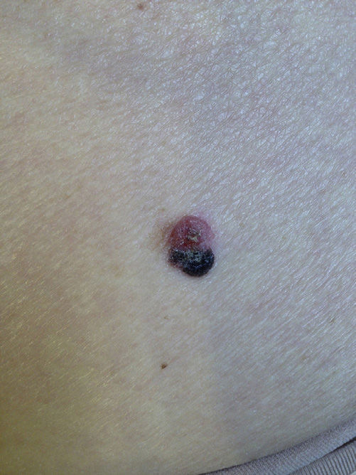
Histologic examination showed hyperkeratosis with parakeratosis, fibrin, and foci of melanin pigment on the surface. There were thick, well-delimited ridges in the Malpighian layer, with no invasion of the underlying dermis. Differentiated keratinocytes, anisocytosis, anisokaryosis, and large hyperchromatic nuclei were observed throughout this layer. There were also horn pearls and hyperpigmentation of keratinocytes without associated melanocytic hyperplasia in the pigmented component. Additional findings included a moderate band-like lymphohistiocytic infiltrate with abundant melanophages in the underlying dermis, under the lesional epithelium (Fig. 3).

What Is Your Diagnosis?
DiagnosisPigmented squamous cell carcinoma in situ in a component of a lesion.
Clinical Course and TreatmentWe widened the tumor margins and found no residual tumor in the specimen analyzed. In 2 years of follow-up, there have been no signs of recurrence or new lesions.
CommentsSSC is the second most common malignant skin tumor, but the pigmented variant of this tumor is rare, accounting for just 7% to 25% of all SCCs.1
Pigmented SCC typically presents as pigmented papules or plaques in sun-exposed areas of the head and neck in elderly individuals.1 Our case is interesting not just because the lesion was pigmented, but also because it involved a patient under 70 years of age and was located in a part of the body that had not been damaged by the sun.2 Dermoscopic features of pigmented SCC include a scaling surface, diffuse or homogeneous blue pigmentation and/or irregularly distributed granular structures; the vessels are often not visible because of the pigment.3 Vascular structures can be highly polymorphous, with irregular hairpin vessels, dotted and/or irregular linear vessels, and spiral vessels, surrounded on occasions by a whitish halo. Chung et al.4 recently published a case of SCC in situ with, as in our case, pigmented projections in peripheral sectors that did not converge geometrically towards the center of the lesion. The authors proposed that this finding suggests a possible nonmelanocytic origin, and together with the scaling, a probable diagnosis of SCC. The presence of whitish perivascular halos suggests a high degree of differentiation,5 which was observed in our case.
The pathophysiology of pigmented SCC is uncertain, and it is thought that the tumor cells might produce cytokines and growth factors that favor melanocytic proliferation and colonization. Another possibility is that the tumor originates from stem cells.6
Histology shows typical features of conventional SCC in addition to pigment in the cytoplasm of the keratinocytes or throughout the tumor in the form of dispersed nonneoplastic dendritic melanocytes and melanophages in the surrounding stroma.2
The differential diagnosis should include pigmented actinic keratosis, pigmented Bowen disease, seborrheic keratosis, pigmented basal cell carcinoma, melanoma, and other adnexal tumors (trichoblastoma and pilomatricoma).6
As there have been few reports of pigmented SCC, little is known about the biologic behavior of this tumor. While pigmentation does not have any prognostic value, it can prompt early consultation.1
In conclusion, although an atypical pigmented lesion in an area of the body protected from the sun may typically suggest melanoma, pigmented BCC, or seborrheic keratosis, the presence of scaling, nongeometric pigmented projections, and a whitish perivascular halo should raise suspicion of BCC.
Conflicts of InterestThe authors declare that they have no conflicts of interest.
Please cite this article as: Guebenlian C, Magliano J, Agorio C. Lesión tumoral pigmentada y vascular en piel no fotoexpuesta. Actas Dermosifiliogr. 2016;107:421–422.


