


The patients were phenotypically identical twin girls who were born healthy following a normal delivery. Neither the parents nor the other 3 siblings of the girls had any relevant medical history. The twins were brought to our unit at age 2 years for assessment of hyperkeratotic lesions on the hands, feet, and periarticular areas; there were no abnormalities of the mucosas or skin appendages. The patients’ mother reported that the lesions had first appeared when the girls were 2 months of age and had progressed slowly.
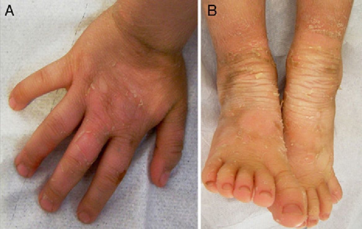
Physical ExaminationBoth girls presented the same clinical picture: thickening and hyperpigmentation of the skin on the hands and feet in a glove-and-stocking distribution as well as in periarticular areas, with superficial scaling in large sheets in some areas. There was also a local increase in fine vellus hair on the limbs (Fig. 1, A and B; Fig. 2, A and B).
Histopathology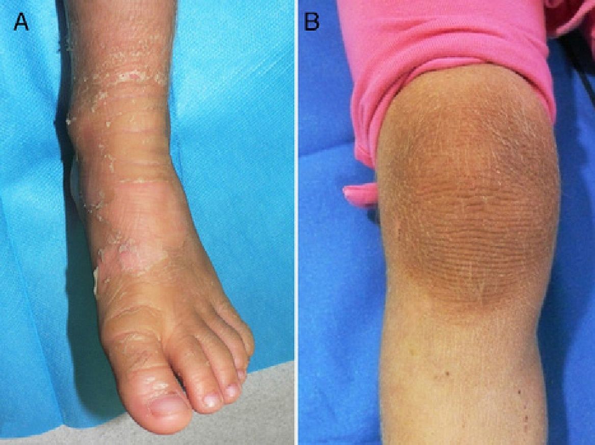
Skin biopsy revealed a mild perivascular inflammatory infiltrate in the upper dermis. The epidermis exhibited eosinophilic orthokeratotic hyperkeratosis with irregular epidermal acanthosis. The stratum granulosum presented intense vacuolization of keratinocytes, loss of intercellular junctions, and epidermal detachment. Large, irregular aggregates of anomalous keratohyalin granules were also observed (Fig. 3).
Additional Tests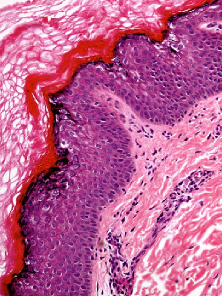
Sequencing of the gene KRT2 (chromosome 12q) revealed a mutation that causes an amino acid change in the protein encoded by this gene.
What Is Your Diagnosis?
DiagnosisIchthyosis bullosa of Siemens (IBS).
Clinical Course and TreatmentBoth patients presented a good response to topical emollient treatment and topical tazarotene (0.03%), and improvement was seen in the treated areas.
CommentIBS is a rare, autosomal dominant disorder that was first described by Siemens1 in 1937. There were no further reports of the condition until 1986, when Traupe et al.2 described a second family affected by the disorder. The underlying genetic alteration in IBS—a mutation in the gene encoding keratin 2e (KRT2e)—was identified in 1994.3 Macroscopically, IBS manifests in periarticular areas (knees, ankles, wrists, and elbows) as areas of superficial epidermal shedding (denuded, apparently healthy skin) surrounded by areas of hyperkeratosis, a sign known as the Mauserung phenomenon.4 This term, coined by Siemens, refers to a distinctive characteristic that is of clinical importance because it is not found in other forms of ichthyosis. Mutations in KRT2e are also expressed in the skin of the palms and soles, but, for reasons unknown, do not cause lesions in those areas.
Mutations in KRT2e are expressed in the stratum granulosum and upper stratum spinosum, where they create a pattern known as epidermolytic hyperkeratosis, characterized microscopically by compact hyperkeratosis and vacuolar degeneration of keratinocytes with alteration of keratohyalin granules. This pattern is not pathognomonic, as it can also be found in other entities such as palmoplantar keratoderma (Vörner type) or epidermolytic acanthoma. Incidental foci of epidermolytic hyperkeratosis can even arise in healthy skin. The main clinical and histologic differential diagnosis of IBS is bullous congenital ichthyosiform erythroderma (CIE), and the 2 conditions are easily confused. Bullous CIE is caused by mutations in the genes encoding keratin 1 or keratin 10, which are expressed deeper in the epidermis, in the suprabasal stratum spinosum. Whereas children with IBS are born healthy, children with bullous CIE are born with erythroderma and present a more severe clinical manifestations, including blister formation, extensive desquamation, and fluid and electrolyte abnormalities.5 A definitive diagnosis of IBS is established by means of genetic testing. Our patients, being monozygotic twins, share the same genetic profile; therefore, they both carry the same mutation. Genetic testing was not performed on the girls’ parents or siblings, as none of them presented phenotypic alterations. Because IBS has an autosomal dominant pattern of inheritance, we hypothesize that our patients developed the disorder as a result of a spontaneous mutation that occurred in the zygote before its initial division. This hypothesis would explain how the mutation was transmitted to both girls. The treatment of IBS is fundamentally supportive, and includes topical emollients and keratolytics. One of the most common complications of this disorder is the development of a secondary infection leading to pustulosis, which requires treatment with topical or systemic antibiotics. IBS is a relatively benign disorder whose clinical manifestations tend to stabilize and improve over time.
Conflicts of InterestThe authors declare that they have no conflicts of interest.
We would like to thank Dr. Antonio Torrelo for his helpful contributions to the diagnosis.
Please cite this article as: Medina-Gil C, et al. Gemelas con cuadro de hiperqueratosis y descamación. Actas Dermosifiliogr. 2012;103:925–6.


